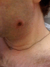
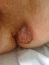
Ulceronodular syphilis (lues maligna praecox) in a person newly diagnosed with HIV infection
- PMID: 22689612
- PMCID: PMC4545021
- DOI: 10.1136/bcr.12.2010.3670
Ulceronodular syphilis (lues maligna praecox) in a person newly diagnosed with HIV infection
Abstract
In this case of secondary syphilis, pustular lesions progressed rapidly to painful ulcerative lesions in a patient with early HIV infection. This rapidly progressive form of early syphilis has historically been called lues maligna praecox, a severe form of noduloulcerative secondary syphilis. Serologic tests for syphilis were positive and biopsy showed forms consistent with Treponema pallidum in the lesions. This case demonstrates how HIV infection may affect presentation and diagnosis of secondary syphilis.
Conflict of interest statement
Figures


References
-
- Fox GH. Cutaneous syphilis. In: Photographic Illustrations of Skin Diseases. New York, NY: E.B. Treat; 1881;53, 65,–68, 81–83.
-
- States WG, Kingsbury J. Syphilis. In: Portfolio of Dermochromes. Volume 3. New York, NY: Rebman Company; 1913;306–8.
-
- Tucker JD, Shah S, Jarell AD, et al. Lues maligna in early HIV infection case report and review of the literature. Sex Transm Dis 2009;36:512–14. - PubMed
-
- Shulkin D, Tripoli L, Abell E. Lues maligna in a patient with human immunodeficiency virus infection. Am J Med 1988;85:425–7. - PubMed
Publication types
MeSH terms
Supplementary concepts
LinkOut - more resources
Full Text Sources
Medical
