. 2012 Nov;97(11):1686-94.
doi: 10.3324/haematol.2011.061408. Epub 2012 Jun 11.

Anti-CD20 IgA can protect mice against lymphoma development: evaluation of the direct impact of IgA and cytotoxic effector recruitment on CD20 target cells

Affiliations

Anti-CD20 IgA can protect mice against lymphoma development: evaluation of the direct impact of IgA and cytotoxic effector recruitment on CD20 target cells

Virginie Pascal et al. Haematologica. 2012 Nov.

Abstract

Background: While most antibody-based therapies use IgG because of their well-known biological properties, some functional limitations of these antibodies call for the development of derivatives with other therapeutic functions. Although less abundant than IgG in serum, IgA is the most abundantly produced Ig class in humans. Besides the specific targeting of its dimeric form to mucosal areas, IgA was shown to recruit polymorphonuclear neutrophils against certain targets more efficiently than does IgG1.

Design and methods: In this study, we investigated the various pathways by which anti-tumor effects can be mediated by anti-CD20 IgA against lymphoma cells.

Results: We found that polymeric human IgA was significantly more effective than human IgG1 in mediating direct killing or growth inhibition of target cells in the absence of complement. We also demonstrated that this direct killing was able to indirectly induce the classical pathway of the complement cascade although to a lesser extent than direct recruitment of complement by IgG. Recruitment of the alternative complement pathway by specific IgA was also observed. In addition to activating complement for lysis of lymphoma cell lines or primary cells from patients with lymphoma, we showed that monomeric anti-CD20 IgA can effectively protect mice against tumor development in a passive immunization strategy and we demonstrated that this protective effect may be enhanced in mice expressing the human FcαRI receptor on their neutrophils.

Conclusions: We show that anti-CD20 IgA antibodies have original therapeutic properties against lymphoma cells, with strong direct effects, ability to recruit neutrophils for cell cytotoxicity and even recruitment of complement, although largely through an indirect way.


Figures

Figure 1.
Figure 1.
Direct effects of anti-CD20 monoclonal antibodies on human lymphoma B cells. Control IgA2 is an anti-βgal mIgA2. (A) Growthinhibitory effect on lymphoma cell lines was measured by the MTS assay. After 48 h with 10 μg of anti-CD20 antibodies, amounts of viable cells were measured for three lymphoma cell lines. (B) Growth inhibition of DHL4 at 48 h was analyzed in the presence of serial dilutions of CD20 antibodies. (C) Apoptosis induction. After 5 h with 10 or 1 μg/mL of each anti-CD20 antibody or control, DHL-4 cells were stained with annexin V-FITC and propidium iodide (PI). The graph shows the percentages of early (Ann+/PI) or late (Ann+/PI+) apoptotic cells. (D) DHL- 4 cells were cultured with 1 μg/mL of anti-CD20 IgG1, pIgA2 or control. At 24 h, BrdU incorporation vs. DNA content (PI) showed the percentage of cells in G0/G1 (BrDU/PIn), S (BrDU+), or G2/M phases (BrDU-/PI2n). Data are presented as mean ± SEM of four independent experiments (Asterisks indicate statistically significant differences between values observed for each form of IgA and IgG1 with an unpaired t-test : *P<0.05, **P<0.01, *** P<0.001).
Figure 2.
Figure 2.
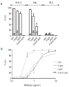
Antibody-complement dependent cytotoxicity against lymphoma cell lines. (A) CDC by anti-CD20 antibodies. DHL-4, BL-2 or Raji cells were incubated with monoclonal antibodies (10 μg/mL) in fresh (open bars) or heat inactivated (gray bars) human serum at 37°C for 4 h. CDC was evaluated by the percentage of propidium iodide-positive cells detected by cell cytometry. (B) Dose-dependence of CDC on DHL-4. The various anti-CD20 monoclonal antibodies were diluted from 10 to 0.1 μg/mL and incubated with DHL-4 for 4 h as above.
Figure 3.
Figure 3.
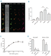
Complement activation by anti-CD20 IgA. (A) Complement depletion studies. DHL-4 cells were incubated with anti-CD20 mIgA2 or IgG1 (10 μg/mL) in normal human serum (NHS), heat inactivated NHS, C1q-depleted serum or factor B-depleted serum at 37°C for 4 h. In control experiments, human exogenous C1q (60 μg/mL) or factor B (300 μg/mL) was added to the depleted serum in order to complement the defect. CDC was evaluated by the percentage of propidium iodide-positive cells by cell cytometry. Data are presented as mean ± SEM of three independent experiments (asterisks indicate statistically significant differences between values tested by an unpaired t test: *P<0.05, **P<0.01). (B) Recruitment of recombinant C1q factor. DHL-4 cells were incubated with 20 μg/mL of anti-CD20-antibodies at 37°C for 2 h. Subsequently, cells were washed and incubated at 4°C for 1 h with 10 μg/mL of recombinant C1q. The attachment of C1q was then revealed by staining with anti-human C1q factor. (C) C1q recruitment and apoptosis. DHL-4 cells were incubated with 20 μg/mL of anti-CD20-antibodies at 37°C for 20 min, 2 h or 5 h. Cells were washed and incubated successively with C1q and anti-human C1q before staining with annexin V (the lower right dot plot appears under-estimated because many cells were completely negative and thus stuck on the horizontal axis).
Figure 4.
Figure 4.
In vivo protective effects of anti-CD20-IgA. (A) RAG2γc-/- were injected intraperitoneally with 106 DHL-4 cells. Various quantities of anti-CD20 or control monoclonal antibodies were also administered i.p. just after cell injection. DHL- 4 specific killing in individual mice was calculated as the ratio of DHL-4 cells and Jurkat reference cells. Data are presented as mean ± SEM of three independent experiments. (B) Human Ig titers were evaluated by ELISA on serum of mice at days 1 and 4 after hydrodynamic injection of plasmid coding for anti-human CD20 antibodies. (C) Western blot analysis of human IgA detected in serum of three different mice 1 day after hydrodynamic tail vein (HTV) injection of pGTRIOIgA2- Ritx-H compared to purified human CD20 IgA in non-reducing conditions with an HRP-linked goat anti-human IgA antibody. (D) Survival curves of C57BL/6 mice (groups of 6 to 7 mice) injected i.v. with 8×10³ EL4-CD20+ cells and treated on day 1 with naked plasmid DNA encoding anti-hCD20 antibodies administered by HTV injection (pGTRIO-IgA2-βgal coding for anti-βgal IgA2, pGTRIO-IgG1-Ritx-H for anti-hCD20 IgG1, pGTRIO-IgA2-Ritx-H for anti-hCD20 IgA2). Asterisks indicate statistically significant differences between survival observed for each form of anti-CD20 antibody and anti-βgal IgA2 treatment with a log-rank test: **P<0.01.
Figure 5.
Figure 5.
Cytotoxic recruitment of CD89+ effector cells by CD20-IgA. (A-C) The ability of anti-CD20 antibodies to mediate human leukocyte recruitment on lymphoma target cells was assessed using the ImageStream® Imaging Flow Cytometer. CFSE-labeled DHL-4 cells were pre-incubated with 20 μg/mL of each CD20 antibody, mixed with human leukocytes and stained with anti-human CD16 PE, anti-human CD45 ECD and DRAQ5 as a nuclear marker. Analyses were performed using the Amnis IDEAS software to evaluate the percentage of CFSE+ targets associated with CD16+ effector leukocytes. (A) Fluorescence and bright field images of representative CFSE+ cells alone or in aggregates with leukocytes. (BF; bright field) (B) Percentage of DHL-4 cells associated with one or more CD16+ effector leukocytes. Asterisks indicate statistically significant differences with a paired t test **P<0.01. (C) Inhibition of mIgA2-mediated target/CD16+ effector aggregation in the presence of antihuman CD89 antibodies. (D) ADCC induced by mIgA2 in human CD89-transgenic or wild-type (WT) mice as evaluated by IVAK; 106 mixed labeled target (EL4-CD20) and reference (EL4 WT) cells were injected i.p. before injection of 20 μg anti-CD20 IgG1, mIgA2 or control. Five hours later, hCD20 -specific killing was calculated from ratios of target to reference cells in the peritoneal washings from individual mice. Data are representative of at least six mice for each condition (asterisks indicate statistically significant differences between values observed for transgenic mice compared to WT mice with an unpaired t test **P<0.01).

References

    1. Ghetie V, Ward ES. Multiple roles for the major histocompatibility complex class Irelated receptor FcRn. Annu Rev Immunol. 2000;18739-66 - PubMed
    1. Bruggemann M, Williams GT, Bindon CI, Clark MR, Walker MR, Jefferis R, et al. Comparison of the effector functions of human immunoglobulins using a matched set of chimeric antibodies. J Exp Med. 1987; 166(5):1351-61 - PMC - PubMed
    1. Pakkanen SH, Kantele JM, Moldoveanu Z, Hedges S, Hakkinen M, Mestecky J, et al. Expression of homing receptors on IgA1 and IgA2 plasmablasts in blood reflects differential distribution of IgA1 and IgA2 in various body fluids. Clin Vaccine Immunol. 2010;17(3):393-401 - PMC - PubMed
    1. Mostov KE. Transepithelial transport of immunoglobulins. Annu Rev Immunol. 1994;1263-84 - PubMed
    1. Daha MR, Gorter A, Rits M, van Es LA, Hiemstra PS. Interaction of immunoglobulin A with complement and phagocytic cells. Prog Clin Biol Res. 1989;297:247-60; discussion 60-1. - PubMed

Publication types

MeSH terms