. 2012 Jul 26;120(4):822-32.
doi: 10.1182/blood-2011-10-387647. Epub 2012 Jun 11.

E6AP ubiquitin ligase regulates PML-induced senescence in Myc-driven lymphomagenesis

Affiliations

E6AP ubiquitin ligase regulates PML-induced senescence in Myc-driven lymphomagenesis

Kamil Wolyniec et al. Blood. .

Erratum in

  • Blood. 2014 Aug 14;124(7):1200

Abstract

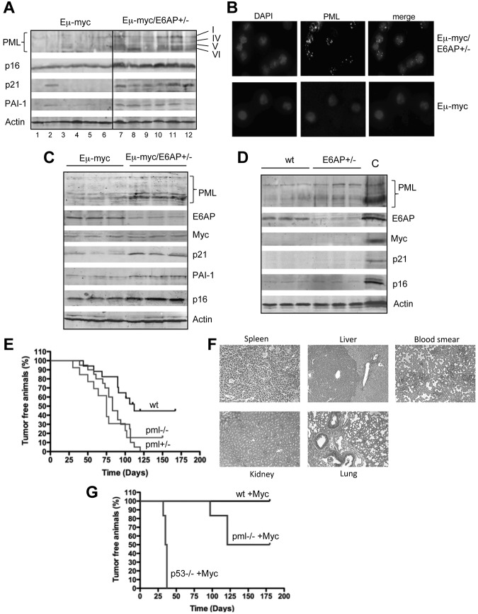
Neoplastic transformation requires the elimination of key tumor suppressors, which may result from E3 ligase-mediated proteasomal degradation. We previously demonstrated a key role for the E3 ubiquitin ligase E6AP in the regulation of promyelocytic leukemia protein (PML) stability and formation of PML nuclear bodies. Here, we report the involvement of the E6AP-PML axis in B-cell lymphoma development. A partial loss of E6AP attenuated Myc-induced B-cell lymphomagenesis. This tumor suppressive action was achieved by the induction of cellular senescence. B-cell lymphomas deficient for E6AP expressed elevated levels of PML and PML-nuclear bodies with a concomitant increase in markers of cellular senescence, including p21, H3K9me3, and p16. Consistently, PML deficiency accelerated the rate of Myc-induced B-cell lymphomagenesis. Importantly, E6AP expression was elevated in ∼ 60% of human Burkitt lymphomas, and down-regulation of E6AP in B-lymphoma cells restored PML expression with a concurrent induction of cellular senescence in these cells. Our findings demonstrate that E6AP-mediated down-regulation of PML-induced senescence is essential for B-cell lymphoma progression. This provides a molecular explanation for the down-regulation of PML observed in non-Hodgkin lymphomas, thereby suggesting a novel therapeutic approach for restoration of tumor suppression in B-cell lymphoma.


Figures

Figure 1
Figure 1
Myc-induced lymphomagenesis is significantly delayed in E6AP heterozygous mice. (A) Death of electronically gated B cells from BM of control wild-type (WT; E6AP+/+) and E6AP−/− mice, which had been irradiated with 5 Gy γ-irradiation was measured by PI staining and analyzed by FACS (n = 3, P < .001). Values represent mean ± SD. (B) Kaplan-Meier survival curves of Eμ-myc/E6AP+/− (n = 36, median survival, 153 days) and control (E6AP+/+) Eμ-myc transgenic mice (n = 30, median survival 103 days). Nontransgenic E6AP+/− mice were used as an additional control (n = 30). Lymphomas developed significantly later in Eμ-myc/E6AP+/− animals compared with the control Eμ-myc mice (P < .001; log-rank test). (C) Numbers of total leukocytes in the blood of sick, lymphoma-burdened mice (n = 15/genotype; P < .01). (D) Weights of spleens of sick, lymphoma burdened mice of the indicated genotypes (n = 15/genotype; P < .001). Values represent mean ± SEM. (E) Weights of combined lymph nodes of sick, lymphoma-burdened mice of the indicated genotypes (n = 15/genotype; P < .01).
Figure 2
Figure 2
The majority of lymphomas derived from Eμ-myc/E6AP+/− mice display a mature sIg+ B cell immunophenotype. (A) Representative dot plots of the most common immunophenotypic profiles obtained by flow cytometric analyses of Eμ-myc/E6AP+/− and control (E6AP+/+) Eμ-myc lymphomas stained with the antibodies to B220, IgM, and IgD. (B) Summary of immunophenotypic analysis of Eμ-myc/E6AP+/− and control (E6AP+/+) Eμ-myc lymphomas.
Figure 3
Figure 3
Preleukemic Eμ-myc/E6AP+/− mice have abnormally reduced numbers of pro-B/pre-B cells but normal numbers of sIg+ B cells compared with control Eμ-myc mice. Cells extracted from the BM and spleens of 4-week-old WT (nontransgenic E6AP+/+; ie, WT), E6AP+/−, Eμ-myc, and Eμ-myc/E6AP+/− mice were stained with antibodies to B220, IgM, and IgD and analyzed by FACS. (A) Absolute numbers of total B cells (B220+; *P < .001), mature B cells (IgM+IgD+; **P < .001), and pro-B/pre-B cells (sIg-B220+; ***P < .001) in BM are presented (n = 3/genotype). Values represent mean ± SEM. (B) Absolute numbers of total B cells (B220+; *P < .001), mature B cells (IgM+IgD+; **P < .001), and pro-B/pre-B cells (sIg-B220+; ***P < .001) in the spleen are presented (n = 3/genotype). Values represent mean ± SEM; NS indicates not statistically significant.
Figure 4
Figure 4
Growth arrest but not increased cell death is evident in lymphoma cells and premalignant B-lymphoid cells from Eμ-myc/E6AP+/− mice. (A) Spontaneous death of Eμ-myc/E6AP+/− and control (E6AP+/+) Eμ-myc lymphoma cells was assayed by staining with PI followed by FACS analysis (n = 10/genotype). Values represent means ± SD. (B) Spontaneous death of BM derived premalignant B-lymphoid cells from 4-week-old Eμ-myc/E6AP+/− and control (E6AP+/+) Eμ-myc mice was assayed by staining with PI followed by FACS analysis (n = 3/genotype). Values represent mean ± SD. (C) Cell-cycle analysis of Eμ-myc/E6AP+/− and control (E6AP+/+) Eμ-myc lymphoma cells showing percentages of cells in each of the phases of cell cycle (n = 10/genotype; *P < .001, **P < .01). Values represent mean ± SD. (D) Cell-cycle analysis of BM derived premalignant B-lymphoid cells of 4-week-old Eμ-myc/E6AP+/− and control (E6AP+/+) Eμ-myc mice was performed by staining with PI, showing percentages of cells in each of the phases of cell cycle (n = 3/genotype; *P < .001; **P < .01). Values represent mean ± SD.
Figure 5
Figure 5
Enhanced cellular senescence in lymphomas and premalignant B-lymphoid cells from Eμ-myc/E6AP+/− mice. (A) Senescence-associated β-galactosidase (SA-β-gal) activity was assayed in lymph nodes and spleen sections at manifestation of lymphoma in Eμ-myc/E6AP+/− and control (E6AP+/+) Eμ-myc mice. Quantification of SA-β-gal positive cells: > 400 cells/tumor, n = 4/genotype were counted in 4 randomly selected fields; *P < .001,**P < .001. Values represent mean ± SD. Magnification ×200. (B) SA-β-gal activity was assayed in cytospins of premalignant B cells isolated from BM of 4-week-old Eμ-myc/E6AP+/− and control (E6AP+/+) Eμ-myc mice. Quantification of SA-β-gal positive cells: > 400 cells/mice, n = 3/genotype were counted in 4 randomly selected fields; P < .001. Values represent means ± SD. Magnification ×200. (C) The levels of H3K9me3 in Eμ-myc/E6AP+/− and control (E6AP+/+) Eμ-myc lymphomas (n = 10/genotype) were measured by FACS analysis after staining with anti-H3K9me3 antibodies. Representative histograms are shown. Summary of results is shown in supplemental Figure 5. (D) The levels of H3K9me3 in BM derived premalignant B lymphoid cells of 4-week-old Eμ-myc/E6AP+/− and control (E6AP+/+) Eμ-myc mice were measured by flow cytometry after staining with anti-H3K9me3 antibodies (n = 3/genotype). Representative histograms are shown.
Figure 6
Figure 6
The critical role for PML in suppression of Myc-driven lymphomas: elevation of PML and PML-NB in lymphoma cells and premalignant B-lymphoid cells from Eμ-myc/E6AP+/− mice. (A) Immunoblot analysis of the indicated proteins in extracts from Eμ-myc/E6AP+/− and control (E6AP+/+) Eμ-myc lymphomas. Probing for actin was used as a loading control. PML isoforms are indicated. (B) Immunofluorescence staining of PML nuclear bodies in lymphomas from Eμ-myc/E6AP+/− and control (E6AP+/+) Eμ-myc mice. Magnification ×1000. (C) Immunoblot analyses of the indicated proteins in premalignant B-lymphoid cells from 4-week-old Eμ-myc/E6AP+/− and control (E6AP+/+) Eμ-myc mice. Probing for actin was used as a loading control. (D) Immunoblot analyses of the indicated proteins in B lymphoid cells from 4-week-old E6AP+/− and control WT (E6AP+/+) mice. Probing for actin was used as a loading control. Lymphoma extract (A, lane 11) was used as a positive control (C). (E) PML loss accelerates tumor onset in Myc-expressing tumors. Eμ-myc transgenic mice were crossed to PML−/− mice. The resulting Eμ-myc mice that were either WT, Pml+/−, or Pml−/− were monitored for tumor onset by spleen as well as lymph node palpation and weekly blood smear analysis. The onset of lymphomas in both Eμ-myc/Pml +/− and Eμ–myc/Pml −/− mice was substantially accelerated compared with control Eμ-myc mice (P < .01). (F) Staining of tissue sections with hematoxylin and eosin revealed that Eμ-myc/Pml−/− lymphomas were highly invasive and infiltrated into the liver, but not kindey or lung. Magnification ×200. (G). HSCs derived from fetal livers of WT, p53−/−, and Pml−/− mice were retrovirally transduced with a MSCV-Myc construct coexpressing green fluorescent protein. The genetically modified stem cells were then used to reconstitute the hematopoietic system of lethally irradiated recipient (WT) animals, which were monitored for lymphoma onset by palpation, weekly blood smears, and whole body fluorescence imaging. Recipients of WT stem cells transduced with the Myc expression did not develop tumors over the observation period, whereas all of the recipients of Myc expression construct transduced p53−/− HSC developed very aggressive disease in a short time. Recipients of Myc expression construct transduced Pml−/− HSCs developed lymphoma more rapidly compared with the mice transplanted with the Myc expression construct transduced WT HSCs (P < .0003), again suggesting that PML can suppress Myc driven lymphoma development.
Figure 7
Figure 7
E6AP levels are elevated in human Burkitt lymphomas and down-regulation of E6AP restores PML-induced cellular senescence. (A) Representative images of E6AP (left panel) and PML (right panel) immunostaining in human Burkitt lymphomas. Expression of E6AP is relatively low in normal lymphoid tissue, but the infiltrating Burkitt lymphoma cells show elevated levels of E6AP accompanied by reduced levels of PML; N = 20; Magnification ×200. (B) Immunoblot analysis to determine the levels of E6AP, PML, and c-Myc in a panel of cell lines derived from Burkitt lymphoma derived (J-1, LOUCIF, LY-67, BL-2, MC116, LS-92, LOUCIF-BL, JOY-BL), DLBCL (SUDHL-4, FARAGE, TOLEDO, DB, HT), or follicular lymphoma (DOHH2). Probing for actin was used as a loading control. The expression levels of E6AP and PML normalized against the levels of actin were quantified and are presented on the graphs (bottom panel). (C) Immunoblot analysis of the indicated proteins in J-1 and LS-92 Burkitt lymphoma derived cells transduced with inducible lentiviral constructs containing wobble E6AP or shRNA for E6AP that were treated with (+) or without (−) doxycyclin. Cells were analyzed 7 days after doxycyclin (dox) induction. Probing for actin was used as a loading control. (D) Immunofluorescence staining of PML-NBs in J-1 Burkitt lymphoma cells transduced with the aforementioned shRNA expression constructs on day 7 after dox treatment. Magnification ×1000. (E) Cell-cycle analyses of J-1 Burkitt lymphoma cells transduced with the aforementioned shRNA expression constructs on performed on day 7 after dox induction; *P < .001. Values represent means ± SD. (F) SA-β-gal levels were determined in cytospins of J-1 Burkitt lymphoma cells transduced with the aforementioned shRNA expression constructs on day 7 after dox treatment. Magnification ×400. Quantification of SA-β-gal positive cells: > 400 cells/cytospin, n = 3, cells were counted in 4 randomly selected fields. Values represent means ± SD, and were derived from 3 independent experiments performed in triplicate.

References

    1. Potts BC, Albitar MX, Anderson KC, et al. Marizomib, a proteasome inhibitor for all seasons: preclinical profile and a framework for clinical trials. Curr Cancer Drug Targets. 2011;11(3):254–284. - PMC - PubMed
    1. Adams J, Kauffman M. Development of the proteasome inhibitor Velcade (Bortezomib). Cancer Invest. 2004;22(2):304–311. - PubMed
    1. Castéra L, Sabbagh A, Dehainault C, et al. MDM2 as a modifier gene in retinoblastoma. J Natl Cancer Inst. 2010;102(23):1805–1808. - PubMed
    1. Leach FS, Tokino T, Meltzer P, et al. p53 Mutation and MDM2 amplification in human soft tissue sarcomas. Cancer Res. 1993;53(10 Suppl):2231–2234. - PubMed
    1. Oliner JD, Kinzler KW, Meltzer PS, George DL, Vogelstein B. Amplification of a gene encoding a p53-associated protein in human sarcomas. Nature. 1992;358(6381):80–83. - PubMed

Publication types

MeSH terms