. 2012 Jul 3;109(27):E1820-9.
doi: 10.1073/pnas.1207829109. Epub 2012 Jun 11.

Infection-induced colitis in mice causes dynamic and tissue-specific changes in stress response and DNA damage leading to colon cancer

Affiliations

Infection-induced colitis in mice causes dynamic and tissue-specific changes in stress response and DNA damage leading to colon cancer

Aswin Mangerich et al. Proc Natl Acad Sci U S A. .

Abstract

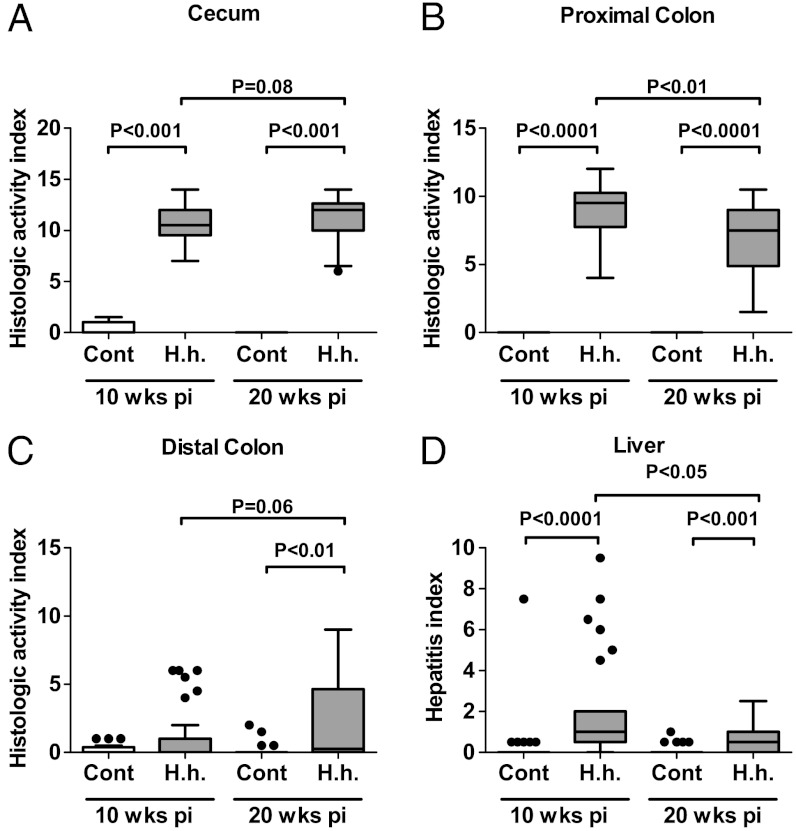
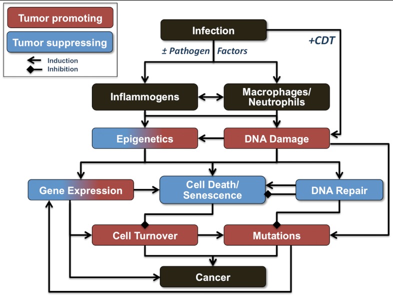
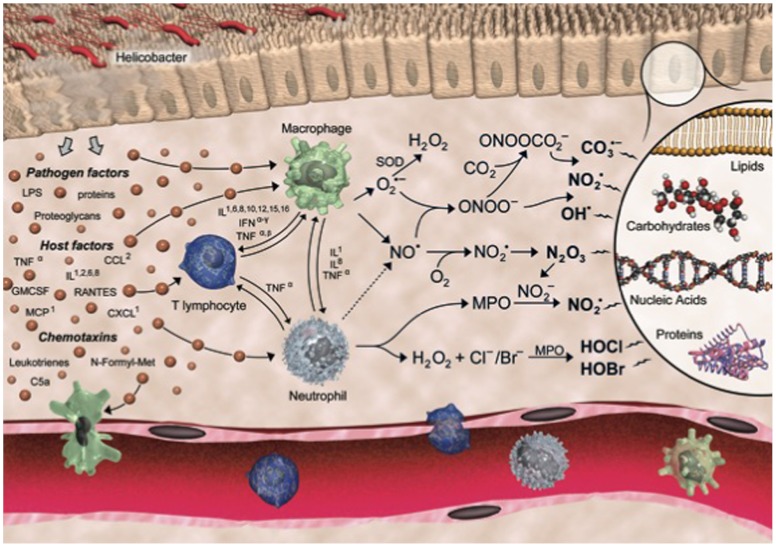
Helicobacter hepaticus-infected Rag2(-/-) mice emulate many aspects of human inflammatory bowel disease, including the development of colitis and colon cancer. To elucidate mechanisms of inflammation-induced carcinogenesis, we undertook a comprehensive analysis of histopathology, molecular damage, and gene expression changes during disease progression in these mice. Infected mice developed severe colitis and hepatitis by 10 wk post-infection, progressing into colon carcinoma by 20 wk post-infection, with pronounced pathology in the cecum and proximal colon marked by infiltration of neutrophils and macrophages. Transcriptional profiling revealed decreased expression of DNA repair and oxidative stress response genes in colon, but not in liver. Mass spectrometric analysis revealed higher levels of DNA and RNA damage products in liver compared to colon and infection-induced increases in 5-chlorocytosine in DNA and RNA and hypoxanthine in DNA. Paradoxically, infection was associated with decreased levels of DNA etheno adducts. Levels of nucleic acid damage from the same chemical class were strongly correlated in both liver and colon. The results support a model of inflammation-mediated carcinogenesis involving infiltration of phagocytes and generation of reactive species that cause local molecular damage leading to cell dysfunction, mutation, and cell death. There are strong correlations among histopathology, phagocyte infiltration, and damage chemistry that suggest a major role for neutrophils in inflammation-associated cancer progression. Further, paradoxical changes in nucleic acid damage were observed in tissue- and chemistry-specific patterns. The results also reveal features of cell stress response that point to microbial pathophysiology and mechanisms of cell senescence as important mechanistic links to cancer.


Conflict of interest statement

The authors declare no conflict of interest.

Figures

Fig. 1.
Fig. 1.
H. hepaticus-infected mice develop chronic colitis, colon carcinoma, and sporadic hepatitis. (A–C) HAIs of colitis were calculated by summing individual scores of inflammation, edema, epithelial defects, crypt atrophy, hyperplasia, and dysplasia for (A) cecum, (B) proximal colon, and (C) distal colon. (D) HIs were calculated by summing individual subfeature histopathology scores from lobular, portal, and interface inflammation, and the number of lobes that contained ≥5 inflammatory lesions. Statistical analysis for all panels is presented as box plots showing the median value (line), the interquartile range (box), and Tukey whiskers embracing data within 1.5-fold of the interquartile range; all data outside the range of the Tukey whiskers are presented as individual data points.
Fig. 2.
Fig. 2.
H. hepaticus-induced colitis is associated tissue infiltration of neutrophils and macrophages. Cell counts (in situ) for MPO-positive neutrophils (A, B) and F4/80-positive macrophages (C, D) in the most affected regions of the lower bowel: cecum (A, C) and proximal colon (B, D). Values represent means ± SD.
Fig. 3.
Fig. 3.
LC-MS/MS quantification of basal levels of nucleobase damage in colon and liver of uninfected mice. Basal levels of nucleobase damage products in DNA and RNA isolated from liver and colon from uninfected Rag2-/- mice. Analyses were performed as described in Materials and Methods. Statistical analysis is presented in box plots as described in Fig. 1.
Fig. 4.
Fig. 4.
H. hepaticus-induced DNA and RNA damage in colon. Rag2-/- mice were infected with H. hepaticus (H.h.) and colon tissue was isolated at 10 and 20 w p.i. for DNA and RNA damage analyses, as described in Materials and Methods. The figure contains data for damage products showing largest increases or decreases, with the complete dataset shown in SI Appendix, Tables S2 and S3. Statistical analysis is presented in box plots as described in Fig. 1.
Fig. 5.
Fig. 5.
Gene expression analysis of DNA repair and oxidative stress response factors in colon and liver tissues of H. hepaticus-infected Rag2-/- mice. QPCR-based gene expression profiling was performed on colon (A, C) and liver (B, D) tissues from control and H. hepaticus-infected mice (20 w p.i.), including 95 key genes involved in generation and detoxification of reactive chemical species (A, B) and DNA damage response (C, D) (SI Appendix, Table S8 lists all genes and gene ontology categories). The data are presented as volcano plots of changes in gene expression levels. Horizontal lines indicate P = 0.05 for N≥5. Vertical lines indicate no change in gene expression. Genes that are significantly up- or down-regulated are labeled with gene symbols.
Fig. 6.
Fig. 6.
Model summarizing the potential mechanisms of inflammation-induced carcinogenesis.
Fig. P1.
Fig. P1.
Chemical biology of Helicobacter-induced inflammation. CCL, chemokine ligand; CXCL, chemokine (C-X-C motif) ligand 1; GMCSF, granulocyte macrophage colony stimulating factor; IFN, interferon; IL, interleukin; LPS, lipopolysaccharide; MCP, monocyte chemotactic protein; MPO, myeloperoxidase; RANTES, regulated upon activation, normal T-cell expressed and secreted; SOD, superoxide dismutase; TNF, tumor necrosis factor.

References

    1. Grivennikov SI, Greten FR, Karin M. Immunity, inflammation, and cancer. Cell. 2010;140:883–899. - PMC - PubMed
    1. Lonkar P, Dedon PC. Reactive species and DNA damage in chronic inflammation: Reconciling chemical mechanisms and biological fates. Int J Cancer. 2011;128:1999–2009. - PMC - PubMed
    1. Dale DC, Boxer L, Liles WC. The phagocytes: Neutrophils and monocytes. Blood. 2008;112:935–945. - PubMed
    1. Terzic J, Grivennikov S, Karin E, Karin M. Inflammation and colon cancer. Gastroenterology. 2010;138:2101–2114. - PubMed
    1. Dedon PC, Tannenbaum SR. Reactive nitrogen species in the chemical biology of inflammation. Arch Biochem Biophys. 2004;423:12–22. - PubMed

Publication types

MeSH terms