. 2012 Jun 26;109(26):10593-8.
doi: 10.1073/pnas.1116590109. Epub 2012 Jun 11.

Reconstitution of abscisic acid activation of SLAC1 anion channel by CPK6 and OST1 kinases and branched ABI1 PP2C phosphatase action

Affiliations

Reconstitution of abscisic acid activation of SLAC1 anion channel by CPK6 and OST1 kinases and branched ABI1 PP2C phosphatase action

Benjamin Brandt et al. Proc Natl Acad Sci U S A. .

Abstract

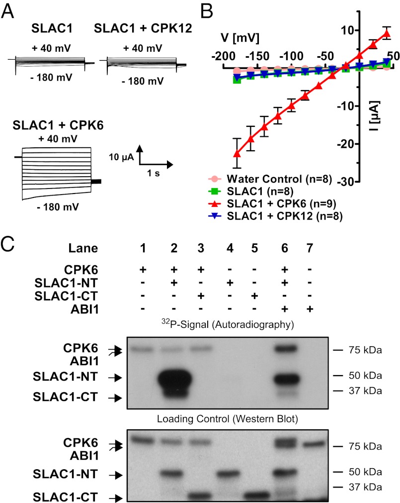
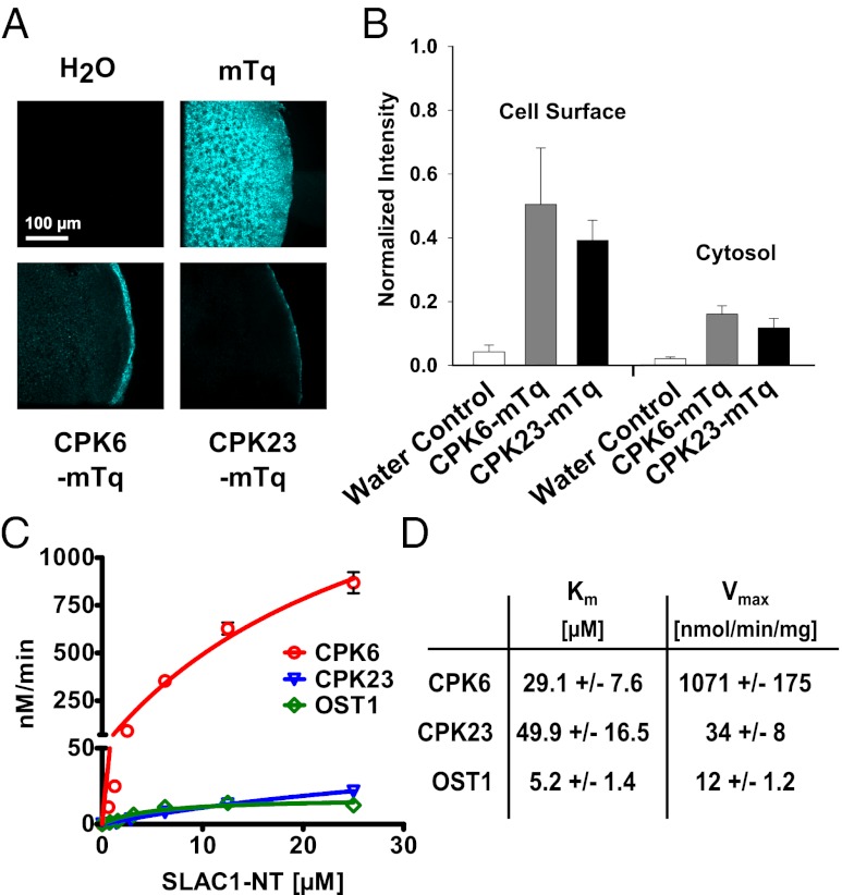
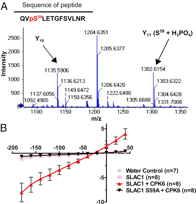
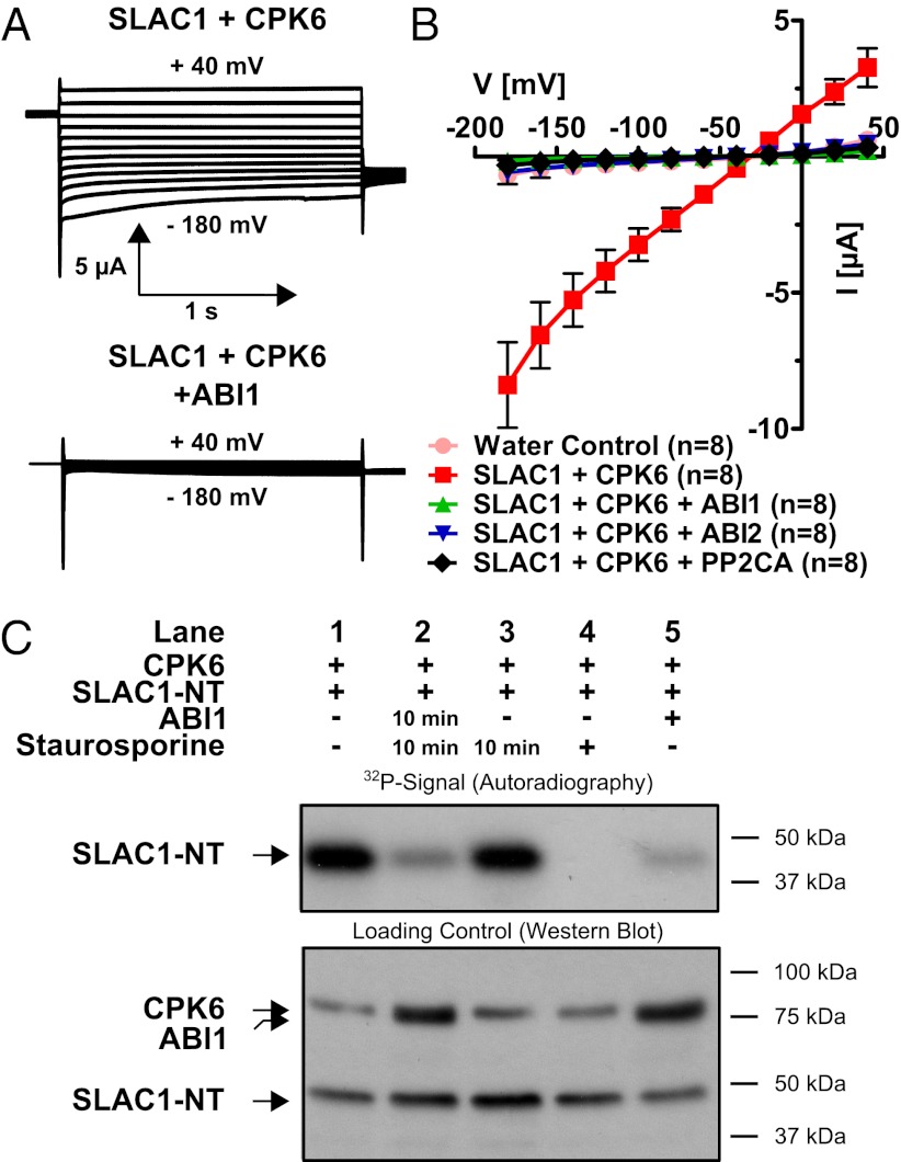
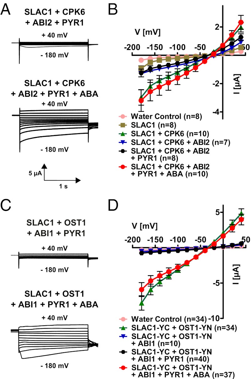
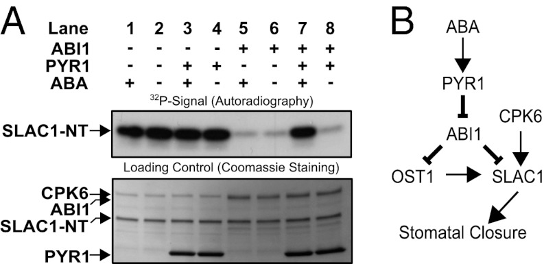
The plant hormone abscisic acid (ABA) is produced in response to abiotic stresses and mediates stomatal closure in response to drought via recently identified ABA receptors (pyrabactin resistance/regulatory component of ABA receptor; PYR/RCAR). SLAC1 encodes a central guard cell S-type anion channel that mediates ABA-induced stomatal closure. Coexpression of the calcium-dependent protein kinase 21 (CPK21), CPK23, or the Open Stomata 1 kinase (OST1) activates SLAC1 anion currents. However, reconstitution of ABA activation of any plant ion channel has not yet been attained. Whether the known core ABA signaling components are sufficient for ABA activation of SLAC1 anion channels or whether additional components are required remains unknown. The Ca(2+)-dependent protein kinase CPK6 is known to function in vivo in ABA-induced stomatal closure. Here we show that CPK6 robustly activates SLAC1-mediated currents and phosphorylates the SLAC1 N terminus. A phosphorylation site (S59) in SLAC1, crucial for CPK6 activation, was identified. The group A PP2Cs ABI1, ABI2, and PP2CA down-regulated CPK6-mediated SLAC1 activity in oocytes. Unexpectedly, ABI1 directly dephosphorylated the N terminus of SLAC1, indicating an alternate branched early ABA signaling core in which ABI1 targets SLAC1 directly (down-regulation). Furthermore, here we have successfully reconstituted ABA-induced activation of SLAC1 channels in oocytes using the ABA receptor pyrabactin resistant 1 (PYR1) and PP2C phosphatases with two alternate signaling cores including either CPK6 or OST1. Point mutations in ABI1 disrupting PYR1-ABI1 interaction abolished ABA signal transduction. Moreover, by addition of CPK6, a functional ABA signal transduction core from ABA receptors to ion channel activation was reconstituted without a SnRK2 kinase.


Conflict of interest statement

The authors declare no conflict of interest.

Figures

Fig. 1.
Fig. 1.
Strong activation of SLAC1 channels by the CPK6 protein kinase in Xenopus oocytes and in vitro. (A) Whole-cell voltage-clamp recordings for oocytes expressing SLAC1, in the presence or absence of CPKs, show that coexpression of SLAC1 with CPK6 results in large Cl currents, as opposed to SLAC1 with CPK12. (B) The average steady-state current–voltage relationships are shown in water-injected (pink circles), SLAC1-injected (green squares), and SLAC1 + CPK-injected oocytes. The protein kinase CPK6 (red triangles) stimulates SLAC1 activity, whereas CPK12 (blue triangles), a homolog of CPK6, did not increase SLAC1 activity in oocytes. Representative data from one batch of oocytes are shown (of >3 batches tested). Error bars represent SEM. (C) Autoradiograph of in vitro kinase assays using recombinant proteins (Upper) and Western blot analysis detecting GST as loading control for the individual proteins (Lower). The kinase CPK6 (lane 1) is able to transphosphorylate the N terminus of SLAC1 (lane 2; SLAC1-NT) but not the C terminus (lane 3; SLAC1-CT). No signal could be detected upon incubation of SLAC1-NT (lane 4) or SLAC1-CT alone (lane 5). Coincubation of CPK6 with ABI1 decreased SLAC1-NT transphosphorylation (lane 6). Assaying ABI1 alone did not result in a 32P signal (lane 7).
Fig. 2.
Fig. 2.
CPK6 is expressed at the cell surface and high in vitro CPK6 activity toward SLAC1-NT phosphorylation. (A) Kinases CPK6 and CPK23 are localized at the cell surface, whereas mTurquoise (mTq) alone is uniformly spread throughout oocytes. (B) Analysis of fluorescence intensity (normalized to mTq) highlights the strong cell-surface localization of both CPK6 and CPK23 (data points represent mean ± SE; n > 3). (C) P81 filter-based protein kinase assays show kinetics of SLAC1-NT phosphorylation by CPK6 (red circles), CPK23 (blue triangles), and OST1 (green diamonds). Data points represent mean values ± SE (n = 3). (D) Km values are given in μM SLAC1-NT, and maximum specific activities are in nmol PO4 incorporated⋅min−1⋅mg−1 kinase.
Fig. 3.
Fig. 3.
Serine 59 of SLAC1 is crucial for CPK6-mediated activation of the anion channel. (A) Zoomed-in spectrum section of product ion scan for the peptide QVpSLETGFSVLNR [(M + 2H)+ = 765.4]. The fragment ion (Y11) corresponding to phosphorylated serine (S59) is labeled in the figure (Y11 (S59 + H3PO4)). The Y10 fragment is labeled to demonstrate that T62 and S65 are not phosphorylated for this ion. The complete spectrum and statistical parameters are presented in Fig. S4A and Table S1. (B) Average steady-state current–voltage relationships show that the point mutation of serine 59 to alanine (black triangles) abolishes anion channel activity due to CPK6 (red triangles). Representative data from one batch of oocytes are shown (of two batches tested).
Fig. 4.
Fig. 4.
Strong inhibition of CPK6-mediated SLAC1 activation by PP2C protein phosphatases and direct SLAC1 N terminus dephosphorylation by ABI1. (A) Whole-cell current recordings for oocytes show that protein phosphatase ABI1 strongly inactivated CPK6-induced SLAC1 ion channel current. (B) Current–voltage relationships in oocytes containing SLAC1 and CPK6 demonstrate that the PP2Cs ABI1 (green triangles), ABI2 (blue triangles), and PP2CA (black diamonds) are capable of strongly inhibiting CPK6-mediated SLAC1 activity (red squares). Data from one representative batch of oocytes (of >3 batches) are shown. (C) (Upper) 32P phosphorylation of the N terminus of SLAC1. (Lower) Loading controls of the individual proteins CPK6, ABI1, and SLAC1. After initial CPK6 exposure, the strong phosphorylation state of SLAC1-NT phosphorylated by CPK6 (Upper, lane 1) decreases if subsequently coincubated with ABI1 and staurosporine 10 min after CPK6 exposure (lane 2). However, phosphorylation of SLAC1-NT by preexposure to CPK6 is not reduced if the protein kinase inhibitor staurosporine alone is added 10 min after CPK6 exposure (lane 3). If staurosporine is added simultaneously with CPK6, SLAC1-NT transphosphorylation is strongly inhibited (lane 4). The ABI1 protein phosphatase added simultaneously with CPK6 inhibits phosphorylation of SLAC1-NT (lane 5).
Fig. 5.
Fig. 5.
Functional reconstitution of ABA activation of SLAC1 channels in (A and B) CPK6- and (C and D) OST1-containing signaling pathways. Current traces (A and C) show examples of the ABA activation of SLAC1 channel currents in oocytes. An ABA-dependent signaling pathway was reconstituted in oocytes using the protein kinases CPK6 (A and B) and OST1 (C and D). Split YFP (BiFC) was used in the case of SLAC1 and OST1 coexpression (C and D). The PP2C phosphatase ABI2 (A and B) or ABI1 (C and D) is able to inhibit SLAC1 currents (blue triangles in B and D). In the absence of ABA, the ABA receptor PYR1 does not activate SLAC1 currents (black hexagons in B and D). However, in the presence of injected ABA, SLAC1 currents are strongly activated (red circles in B and D). Data from one representative batch of oocytes (of >3 batches) are shown in D. Data from two to five independent batches were averaged in B.
Fig. 6.
Fig. 6.
(A) In vitro reconstitution of an ABA-dependent signal transduction pathway including PYR1, ABI1, CPK6, and SLAC1-NT protein. Independent of ABA, CPK6 phosphorylates SLAC1-NT (lanes 1 and 2). The presence of ABI1, but not PYR1, inhibits SLAC1-NT phosphorylation, with or without ABA (lanes 3–6). If CPK6, ABI1, and PYR1 are present, addition of ABA leads to the release of ABI1-mediated inhibition of SLAC1 phosphorylation (lane 7). (B) Model for ABA activation of the SLAC1 channel via the OST1 and CPK6 protein kinases and down-regulation by the ABI1 protein phosphatase. ABA binds to PYR1, which causes inhibition of PP2C protein phosphatases, including ABI1. This leads to ABA-induced activation of SLAC1 channels in Xenopus oocytes by CPK6, which functions in native guard cells in ABA activation of S-type anion channels (37). Note that the ABI1 protein phosphatase can directly dephosphorylate the SLAC1 N terminus, which represents a previously unknown target for ABI1 and a mechanism for tight negative SLAC1 regulation, in addition to the known down-regulation of OST1.

References

    1. Ma Y, et al. Regulators of PP2C phosphatase activity function as abscisic acid sensors. Science. 2009;324:1064–1068. - PubMed
    1. Park S-Y, et al. Abscisic acid inhibits type 2C protein phosphatases via the PYR/PYL family of START proteins. Science. 2009;324:1068–1071. - PMC - PubMed
    1. Nishimura N, et al. PYR/PYL/RCAR family members are major in-vivo ABI1 protein phosphatase 2C-interacting proteins in Arabidopsis. Plant J. 2010;61:290–299. - PMC - PubMed
    1. Santiago J, et al. Modulation of drought resistance by the abscisic acid receptor PYL5 through inhibition of clade A PP2Cs. Plant J. 2009;60:575–588. - PubMed
    1. Szostkiewicz I, et al. Closely related receptor complexes differ in their ABA selectivity and sensitivity. Plant J. 2010;61(1):25–35. - PubMed

Publication types

MeSH terms

Substances