. 2012 Jun 26;109(26):10310-5.
doi: 10.1073/pnas.1202071109. Epub 2012 Jun 11.

Arylsulfatase G inactivation causes loss of heparan sulfate 3-O-sulfatase activity and mucopolysaccharidosis in mice

Affiliations

Arylsulfatase G inactivation causes loss of heparan sulfate 3-O-sulfatase activity and mucopolysaccharidosis in mice

Björn Kowalewski et al. Proc Natl Acad Sci U S A. .

Abstract

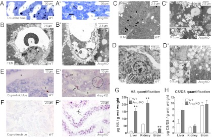
Deficiency of glycosaminoglycan (GAG) degradation causes a subclass of lysosomal storage disorders called mucopolysaccharidoses (MPSs), many of which present with severe neuropathology. Critical steps in the degradation of the GAG heparan sulfate remain enigmatic. Here we show that the lysosomal arylsulfatase G (ARSG) is the long-sought glucosamine-3-O-sulfatase required to complete the degradation of heparan sulfate. Arsg-deficient mice accumulate heparan sulfate in visceral organs and the central nervous system and develop neuronal cell death and behavioral deficits. This accumulated heparan sulfate exhibits unique nonreducing end structures with terminal N-sulfoglucosamine-3-O-sulfate residues, allowing diagnosis of the disorder. Recombinant human ARSG is able to cleave 3-O-sulfate groups from these residues as well as from an authentic 3-O-sulfated N-sulfoglucosamine standard. Our results demonstrate the key role of ARSG in heparan sulfate degradation and strongly suggest that ARSG deficiency represents a unique, as yet unknown form of MPS, which we term MPS IIIE.


Conflict of interest statement

The authors declare no conflict of interest.

Figures

Fig. 1.
Fig. 1.
Neuropathology of Arsg-deficient mice. (A and B) Toluidine blue-stained Epon-embedded sections of the CNS reveal storage vacuoles (arrows) in Purkinje cells (A) and perivascular macrophages (B). (C) Storage vacuoles in both Purkinje cells (long arrow) and macrophages (short arrows) show positive periodic acid–Schiff (PAS) staining. (D) TEM reveals heterogeneous, partially water-soluble, and finely granular to lamellar storage material with variable electron density in Purkinje cells. (E and E′) Fluorescence microscopy of unstained sections shows autofluorescence in the cerebellar cortex of Arsg-deficient mice but not in WT animals. (F and F′) Autofluorescent storage material (red) colocalizes with ring-like structures of the lysosomal membrane protein Lamp1 (green), indicating its lysosomal origin. (G and G′) Considerable loss of Purkinje cells can be observed by 12 mo of age in Arsg-deficient mice as assessed by Calbindin immunohistochemistry. (Inset) Remaining Purkinje cells show swollen dendrites (arrows) and somata (arrowhead). (HJ′) Profound activation of microglia transforming to an amoeboid to phagocytic morphology (H and H, I and I′) and hypertrophy of astrocytes (J and J′) occur in Arsg-deficient animals in the cerebellar cortex as assessed with markers for microglia/macrophages (CD68) and astrocytes (GFAP) (age 12 mo). (K and L) Arsg-deficient mice showed a progressive exploratory deficit in the open-field test as indicated by reduced distance traveled (K) and time spent (L) in the center. (M) Lower step-through latencies indicate impaired passive-avoidance learning in Arsg-deficient mice. (N) Water-maze probe trial shows impaired visuo-spatial memory for the platform location in Arsg-deficient mice. For K–N, n = 20, age 12 mo; error bars indicate SEM.
Fig. 2.
Fig. 2.
Lysosomal storage pathology of liver and kidney. (A and A′) Toluidine blue-stained Epon-embedded sections of liver from Arsg-deficient mice (12 mo old) reveal abnormal vacuoles in the peribiliary cytoplasm of hepatocytes (arrows) (S, sinusoid; LD, lipid droplet). (B and B′) Endothelial cell of hepatic sinusoid contains cytoplasmic vacuoles with remnants of electron-dense material. (C and C′) Cytoplasmic vacuoles in hepatocytes of Arsg-deficient mice. (D and D′) In kidney, the epithelium of the thick ascending limbs of Henle’s loop displays cytoplasmic vacuoles that appear almost empty. (EF′) Cuprolinic blue incubation of liver (E and E′) and kidney (F and F′) sections reveals positive staining of inclusion bodies in hepatocytes (circle) and strong metachromatic GAG staining in liver sinusoidal cells (arrow) and in kidney thick ascending limbs (TAL) of Arsg-deficient mice. (G and H) Quantification of GAGs extracted from liver, brain, and kidney of Arsg-knockout and WT mice. The amounts of heparan sulfate (G) and chondroitin/dermatan sulfate (CS/DS) (H) were determined by LC/MS. Error bars indicate SD (n = 3).
Fig. 3.
Fig. 3.
NRE analysis of heparan sulfate from Arsg-knockout and WT livers. Heparan sulfate was enzymatically depolymerized and analyzed by LC/MS to detect the liberated NRE saccharides. (A) The accumulative extracted ion-current chromatograms for the indicated NRE structures are shown. One monosaccharide (dp1) and four trisaccharides (dp3) were identified as NRE saccharides in heparan sulfate from Arsg-knockout mouse liver (black trace). Little or no NRE structures were observed in WT liver (red trace). (B) Before LC/MS analysis, [13C6]aniline-labeled GlcNS3S (red trace) was added to depolymerized Arsg-knockout heparan sulfate tagged with [12C6]aniline (black trace). The dp1 structure shown in A coelutes with the standard. (C) An equimolar mixture of standard GlcNS3S and GlcNS6S was differentially labeled with [13C6]aniline and [12C6]aniline, respectively, and chromatographically resolved (see molecular structures after aniline labeling at the bottom of the figure). (D) Mass spectrum for the experiment of B showing both [12C6]aniline-labeled NRE monosaccharide (black) and [13C6]aniline-labeled GlcNS3S (red) with the expected 6.02 mass unit difference attributable to differential isotope labeling. (E) Mass spectrum for [13C6]aniline-labeled GlcNS3S, demonstrating only the free molecular ion (m/z = 421.07). (F) Mass spectrum for [12C6]aniline-labeled GlcNS6S, showing both the free molecular ion (m/z = 415.05, [M-H]) and a stable adduction ion (m/z = 544.20, [M-2H+DBA]) formed with the ion-pairing reagent DBA during LC/MS.
Fig. 4.
Fig. 4.
In vitro analysis of ARSG substrate specificity. The activity of purified rhARSG was assayed with various substrates. (A) rhARSG mediates sulfate release from GlcNS3S but not from GlcNS or GlcN3S. rhARSA does not act on any of the substrates. Error bars indicate SEM (n = 3). (B and D) GlcNS3S, but not GlcNS6S, is desulfated to GlcNS upon treatment with ∼10 mU of rhARSG for 16 h (red traces) as determined by LC/MS analysis. Controls were treated with heat-inactivated BSA (black traces). All traces represent accumulative extracted-ion-current chromatograms. (C) Heparan sulfate (250 pmol) from Arsg-knockout liver lysosome-enriched fractions (tritosomes) was subjected to quantitative NRE analysis after treatment with rhARSG (∼2 mU) or heat-inactivated BSA for 16 h. The amount of GlcNS3S NRE is shown for both conditions.
Fig. 5.
Fig. 5.
Heparan sulfate catabolism involving GlcNS3S structures. The scheme illustrates all nine different enzymatic activities required for the sequential catabolism of a NRE tetrasaccharide containing GlcNS3S. To expose the 3-O-sulfated residue at the terminus, the preceding uronic acid (iduronate 2-O-sulfate in this example) is modified sequentially by iduronate 2-sulfatase and iduronidase. Under normal conditions, the 3-O-sulfate then is removed from GlcNS3S by ARSG, thus generating the substrate for sulfamidase, which removes the N-sulfate group. Subsequently, another six different enzymes (plus again sulfamidase) have to act, which ultimately leads to a complete degradation of the chain. The loss of ARSG activity (MPS IIIE) leads to the accumulation of 3-O-sulfated ARSG substrate that cannot be acted upon by downstream catabolic enzymes. It should be noted that the 2-O-sulfation shown at the glucuronic acid (third residue) is relatively rare, which agrees with the finding that no pentasulfated trisaccharides were found as NRE structures (Fig. 3A). Scheme modified from Neufeld and Muenzer (6) according to findings from this work and from Lawrence et al. (12).

References

    1. Ballabio A, Gieselmann V. Lysosomal disorders: From storage to cellular damage. Biochim Biophys Acta. 2009;1793:684–696. - PubMed
    1. Vitner EB, Platt FM, Futerman AH. Common and uncommon pathogenic cascades in lysosomal storage diseases. J Biol Chem. 2010;285:20423–20427. - PMC - PubMed
    1. Diez-Roux G, Ballabio A. Sulfatases and human disease. Annu Rev Genomics Hum Genet. 2005;6:355–379. - PubMed
    1. Mehl E, Jatzkewitz H. Cerebroside 3-sulfate as a physiological substrate of arylsulfatase A. Biochim Biophys Acta. 1968;151:619–627. - PubMed
    1. von Figura K, Gieselmann V, Jaeken J. Metachromatic leukodystrophy. In: Scriver CR, et al., editors. The Metabolic and Molecular Bases of Inherited Diseases. New York: McGraw-Hill; 2001. pp. 3694–3724.

Publication types