

Expression of MALT1 oncogene in hematopoietic stem/progenitor cells recapitulates the pathogenesis of human lymphoma in mice
- PMID: 22689981
- PMCID: PMC3387079
- DOI: 10.1073/pnas.1204127109
Expression of MALT1 oncogene in hematopoietic stem/progenitor cells recapitulates the pathogenesis of human lymphoma in mice
Abstract
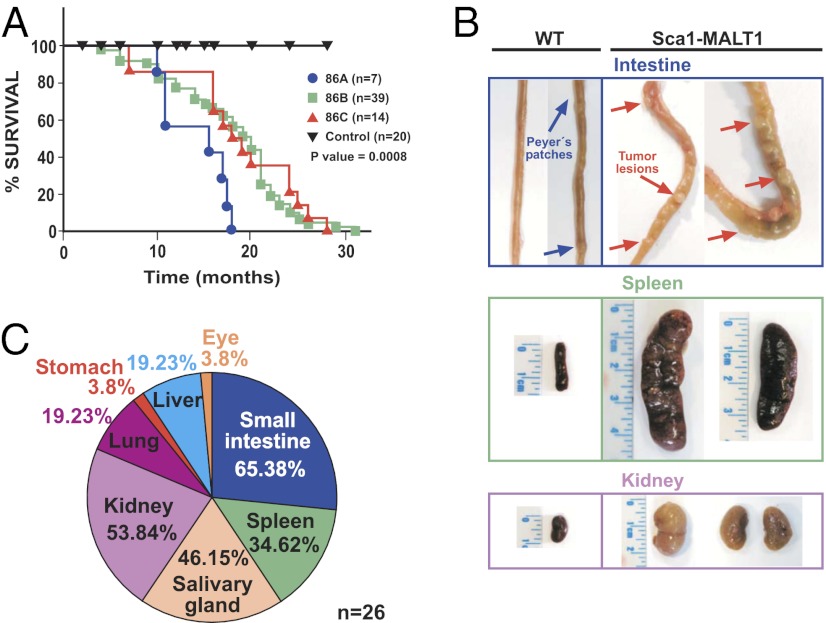
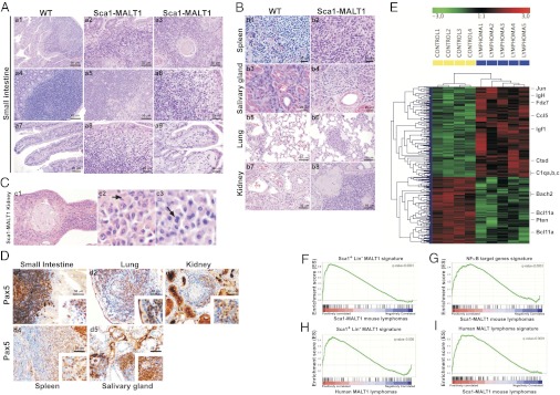
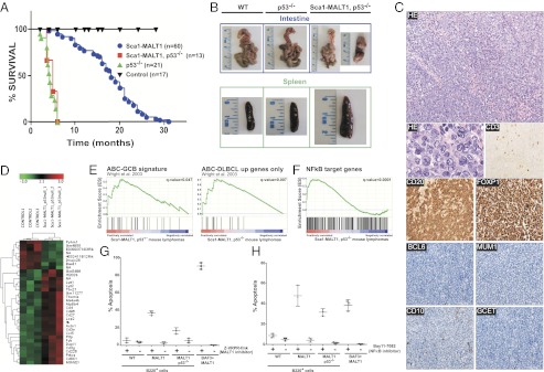
Chromosomal translocations involving the MALT1 gene are hallmarks of mucosa-associated lymphoid tissue (MALT) lymphoma. To date, targeting these translocations to mouse B cells has failed to reproduce human disease. Here, we induced MALT1 expression in mouse Sca1(+)Lin(-) hematopoietic stem/progenitor cells, which showed NF-κB activation and early lymphoid priming, being selectively skewed toward B-cell differentiation. These cells accumulated in extranodal tissues and gave rise to clonal tumors recapitulating the principal clinical, biological, and molecular genetic features of MALT lymphoma. Deletion of p53 gene accelerated tumor onset and induced transformation of MALT lymphoma to activated B-cell diffuse large-cell lymphoma (ABC-DLBCL). Treatment of MALT1-induced lymphomas with a specific inhibitor of MALT1 proteolytic activity decreased cell viability, indicating that endogenous Malt1 signaling was required for tumor cell survival. Our study shows that human-like lymphomas can be modeled in mice by targeting MALT1 expression to hematopoietic stem/progenitor cells, demonstrating the oncogenic role of MALT1 in lymphomagenesis. Furthermore, this work establishes a molecular link between MALT lymphoma and ABC-DLBCL, and provides mouse models to test MALT1 inhibitors. Finally, our results suggest that hematopoietic stem/progenitor cells may be involved in the pathogenesis of human mature B-cell lymphomas.
Conflict of interest statement
The authors declare no conflict of interest.
Figures
References
-
- Farinha P, Gascoyne RD. Molecular pathogenesis of mucosa-associated lymphoid tissue lymphoma. J Clin Oncol. 2005;23:6370–6378. - PubMed
-
- Isaacson PG, Du MQ. MALT lymphoma: From morphology to molecules. Nat Rev Cancer. 2004;4:644–653. - PubMed
-
- Sagaert X, De Wolf-Peeters C, Noels H, Baens M. The pathogenesis of MALT lymphomas: Where do we stand? Leukemia. 2007;21:389–396. - PubMed
-
- Dierlamm J, et al. The apoptosis inhibitor gene API2 and a novel 18q gene, MLT, are recurrently rearranged in the t(11;18)(q21;q21) associated with mucosa-associated lymphoid tissue lymphomas. Blood. 1999;93:3601–3609. - PubMed
-
- Sanchez-Izquierdo D, et al. MALT1 is deregulated by both chromosomal translocation and amplification in B-cell non-Hodgkin lymphoma. Blood. 2003;101:4539–4546. - PubMed
Publication types
MeSH terms
Substances
Associated data
- Actions
- Actions
- Actions
- Actions
- Actions
LinkOut - more resources
Full Text Sources
Other Literature Sources
Medical
Molecular Biology Databases
Research Materials
Miscellaneous
