. 2012 Jun 26;109(26):10516-21.
doi: 10.1073/pnas.1202684109. Epub 2012 Jun 11.

A discrete population of squamocolumnar junction cells implicated in the pathogenesis of cervical cancer

Affiliations

A discrete population of squamocolumnar junction cells implicated in the pathogenesis of cervical cancer

Michael Herfs et al. Proc Natl Acad Sci U S A. .

Abstract

Infection by carcinogenic human papillomaviruses (HPV) results in precancers [cervical intraepithelial neoplasia (CIN)] and cancers near the ectoendocervical squamocolumnar (SC) junction of the cervix. However, the specific cells targeted by HPV have not been identified and the cellular origin of cervical cancer remains elusive. In this study, we uncovered a discrete population of SC junctional cells with unique morphology and gene-expression profile. We also demonstrated that the selected junctional biomarkers were expressed by a high percentage of high-grade CIN and cervical cancers associated with carcinogenic HPVs but rarely in ectocervical/transformation zone CINs or those associated with noncarcinogenic HPVs. That the original SC junction immunophenotype was not regenerated at new SC junctions following excision, not induced by expression of viral oncoproteins in foreskin keratinocytes, and not seen in HPV-related precursors of the vagina, vulva, and penis further support the notion that junctional cells are the source of cervical cancer. Taken together, our findings suggest that carcinogenic HPV-related CINs and cervical cancers are linked to a small, discrete cell population that localizes to the SC junction of the cervix, expresses a unique gene expression signature, and is not regenerated after excision. The findings in this study uncover a potential target for cervical cancer prevention, provide insight into the risk assessment of cervical lesions, and establish a model for elucidating the pathway to cervical cancer following carcinogenic HPV infection.


Conflict of interest statement

The authors declare no conflict of interest.

Figures

Fig. 1.
Fig. 1.
Identification and transcriptional analysis of SC junction cells. (A) Schematic representation of the human cervix. (B) Histology of adult cervix, with squamous (Top), junctional (Middle), and columnar (Bottom) cells. (C) Two-dimensional representation of a principle component analysis of expression microarray data derived from the three cell populations. (D) Heat map of expression microarray data anchored by a comparison between the three groups; SC junction-specific expression is bracketed.
Fig. 2.
Fig. 2.
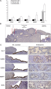
Characterization of several SC junction-specific biomarkers. (A) Fold differences in expression of five genes within the squamous, columnar, and junctional cells. These results are derived from microarray analysis. (B) Low-magnification image showing the entire cervix. Krt7 staining highlights a discrete population of cuboidal cells at the interface of the transformation zone squamous epithelium and endocervix. (C) Antibodies stain an identical cell population at the SC junction. The positive staining extends from the surface of the stratified squamous epithelium onto the adjacent basement membrane. Note the absence of staining of either the mature keratinocytes or the endocervical columnar cells.
Fig. 3.
Fig. 3.
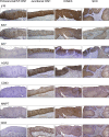
Immunohistochemistry of sections of human cervix with CIN and squamous carcinoma for p16, Ki67, and the five SC junction-specific antibodies. Note the absence of staining of the ectocervical/TZ CIN1s and uniform staining of junctional CINs of all grades and squamous carcinoma. The expression of SC junction-specific markers was assessed in 34 ectocervical/TZ CIN1, 8 junctional CIN1, 48 CIN2/3, and 10 cancers.
Fig. 4.
Fig. 4.
Topographic specificity of the SC junction immunophenotype. (A) Fluorescence micrograph of human cervix at 16 wk (Top) showing diffuse Krt7 immunopositivity. At 20 wk of gestation, basal Krt5 expression emerges (Middle). In the adult cervix (Bottom), the Krt7 staining is limited to the SC junction. For each case, a corresponding histology image [hematoxylin–eosin staining (HE)] is shown. (B) Western blots of lysates of control (−), HPV16 E6- or E7-expressing primary human keratinocyte cultures, cervical adenocarcinoma (HeLa), and squamous carcinoma (SiHa, CaSki) reacted with antibodies specific for the SC junctional cells. Only cervix-derived tumor cells (HeLa, SiHa, and CaSki) score positive. (C) Schematic illustration of the squamocolumnar junction before and after LEEP (Left and Center). Absence of Krt7 staining in a “new” SC junction following LEEP (Right).

Similar articles

Cited by

References

    1. Dürst M, Gissmann L, Ikenberg H, zur Hausen H. A papillomavirus DNA from a cervical carcinoma and its prevalence in cancer biopsy samples from different geographic regions. Proc Natl Acad Sci USA. 1983;80:3812–3815. - PMC - PubMed
    1. Boshart M, et al. A new type of papillomavirus DNA, its presence in genital cancer biopsies and in cell lines derived from cervical cancer. EMBO J. 1984;3:1151–1157. - PMC - PubMed
    1. Crum CP, Ikenberg H, Richart RM, Gissman L. Human papillomavirus type 16 and early cervical neoplasia. N Engl J Med. 1984;310:880–883. - PubMed
    1. Lorincz AT, Lancaster WD, Temple GF. Cloning and characterization of the DNA of a new human papillomavirus from a woman with dysplasia of the uterine cervix. J Virol. 1986;58:225–229. - PMC - PubMed
    1. Richart RM. Cervical intraepithelial neoplasia. Pathol Annu. 1973;8:301–328. - PubMed

Publication types