. 2012 Jun 26;109(26):10474-9.
doi: 10.1073/pnas.1114214109. Epub 2012 Jun 11.

Opposing roles for RhoH GTPase during T-cell migration and activation

Affiliations

Opposing roles for RhoH GTPase during T-cell migration and activation

Christina M Baker et al. Proc Natl Acad Sci U S A. .

Abstract

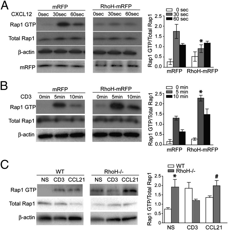
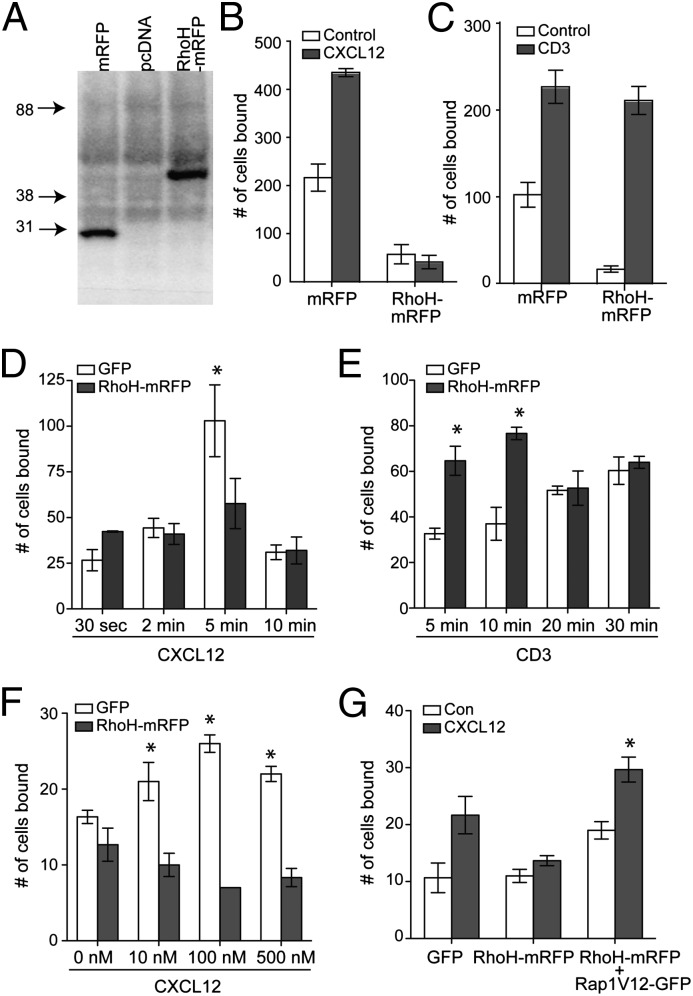
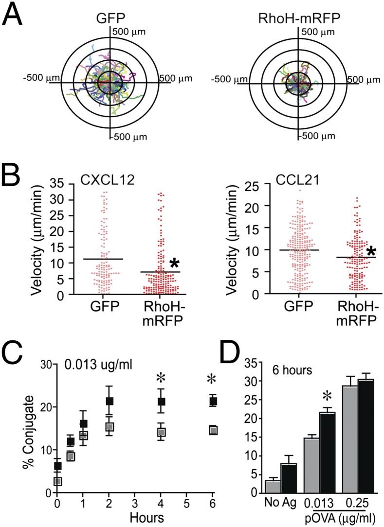
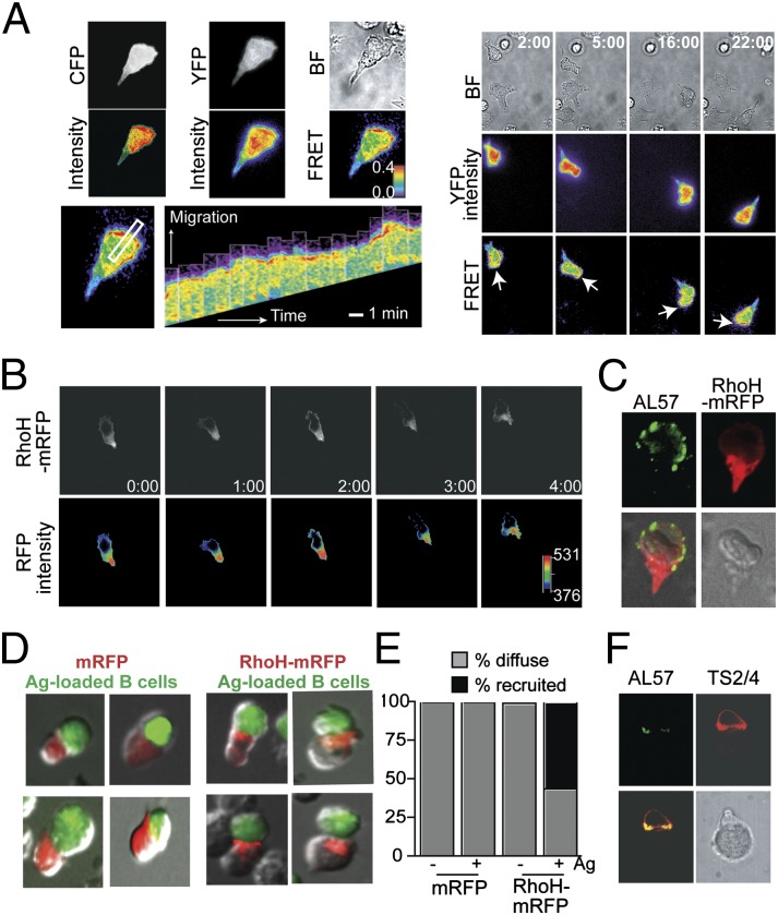
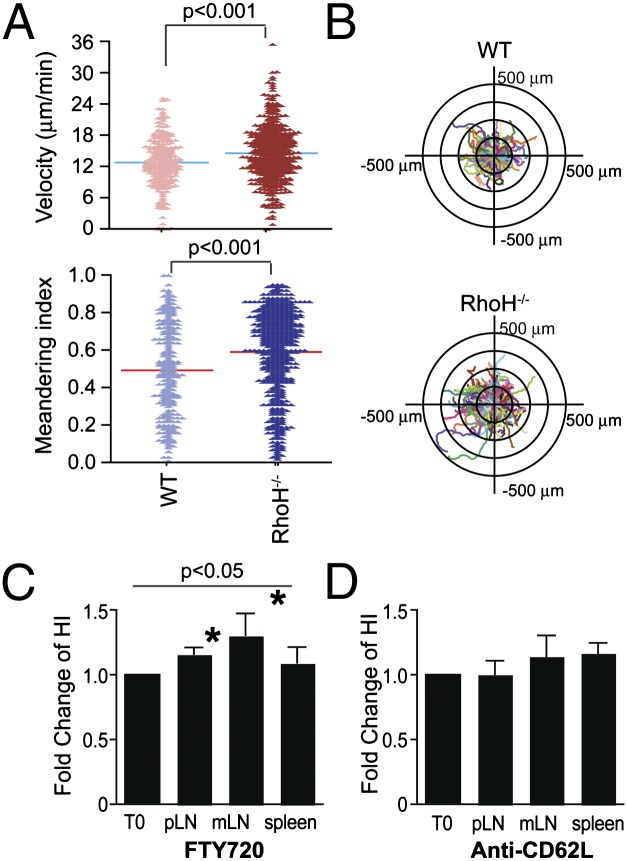
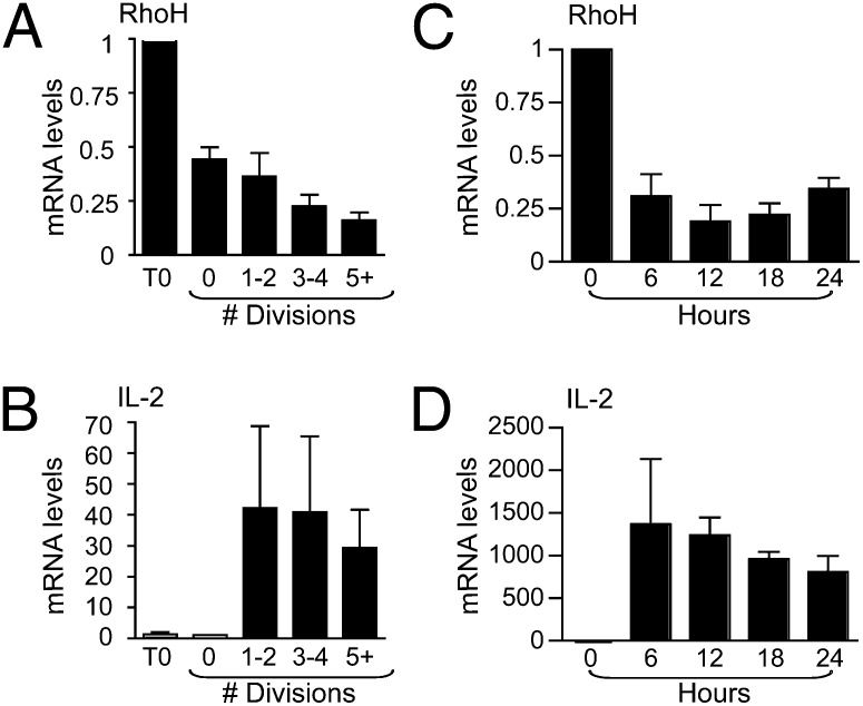
T cells spend the majority of their time perusing lymphoid organs in search of cognate antigen presented by antigen presenting cells (APCs) and then quickly recirculate through the bloodstream to another lymph node. Therefore, regulation of a T-cell response is dependent upon the ability of cells to arrive in the correct location following chemokine gradients ("go" signal) as well as to receive appropriate T-cell receptor (TCR) activation signals upon cognate antigen recognition ("stop" signal). However, the mechanisms by which T cells regulate these go and stop signals remain unclear. We found that overexpression of the hematopoietic-specific RhoH protein in the presence of chemokine signals resulted in decreased Rap1-GTP and LFA-1 adhesiveness to ICAM-1, thus impairing T-cell chemotaxis; while in the presence of TCR signals, there were enhanced and sustained Rap1-GTP and LFA-1 activation as well as prolonged T:APC conjugates. RT-PCR analyses of activated CD4(+) T cells and live images of T-cell migration and immunological synapse (IS) formation revealed that functions of RhoH took place primarily at the levels of transcription and intracellular distribution. Thus, we conclude that RhoH expression provides a key molecular determinant that allows T cells to switch between sensing chemokine-mediated go signals and TCR-dependent stop signals.


Conflict of interest statement

The authors declare no conflict of interest.

Figures

Fig. 1.
Fig. 1.
RhoH negatively regulates chemokine-mediated Rap1 activation and positively regulates TCR-mediated Rap1 activation. (AC) Jurkat cells were transiently transfected with mRFP or RhoH–mRFP, then at various time after stimulation with CXCL12 or CD3 cross-linking, cell lysates were assessed for active Rap1, total Rap-1, and b-actin. Representative blots of Rap1 activation by CXCL12 (A) or CD3 cross-linking (B) are shown and bar graphs of each pair of blots consist of compiled densitometry analysis (Right). (C) Freshly isolated CD4+ T cells from WT and RhoH−/− mice were unstimulated (NS) or stimulated with CD3 cross-linking or CCL21 and Rap1–GTP assessed. Data are means ± SEM, n = 3, and statistically significant, *P < 0.05 vs. WT NS, statistically significant, #P < 0.03 vs. RhoH−/− CD3.
Fig. 2.
Fig. 2.
RhoH negatively regulates chemokine-mediated LFA-1 adhesion and positively regulates TCR-mediated LFA-1 adhesion. (AC) Stable Jurkat cell lines expressing pcDNA, mRFP, or RhoH–mRFP were generated and verified by anti-mRFP immunoblot. Cells were stimulated with CXCL12 (B) or OKT3 and secondary antibody (C) to induce TCR cross-linking, then assessed in an ICAM-1 adhesion assay. (DG) Primary human lymphocytes transiently transfected with GFP or RhoH–mRFP were sorted. ICAM-1 adhesion of CXCL12 stimulation (D) or TCR stimulation (E) over time, CXCL12 titration (F) or ICAM-1 adhesion of CXCL12 stimulation of control, RhoH overexpressing, or double positive RhoH–mRFP and Rap1V12–GFP (G). Data are means ± SEM. For B, n = 4 and P < 0.02 and CG n = 3 independent donors.
Fig. 3.
Fig. 3.
RhoH negatively regulates chemokine-induced T-cell migration and positively regulates sustained T:APC conjugates. (A) Human T-cell blasts were transiently transfected with control vector (GFP) or RhoH–mRFP vector. Cell migration on CXCL12/ICAM-1 was analyzed; corresponding cell tracking plots are shown (A, Movie S1). Images include fluorescent signal to indicate transfected cells overlaid with migration tracks as indicated by colored lines. (B) Velocity (millimeters per minute) of cell migration induced by CXCL12 or CCL21 on ICAM-1 was analyzed. Data are combined from three independent donors and are statistically significant P < 0.001 by Mann–Whitney test. (C and D) DO11.10 TCR transgenic T-cell blasts were transiently transfected with control vector or RhoH–mRFP and assessed in a flow cytometry-based conjugate assay. (C) Representative plot of conjugate formation over time at one antigen dose. (D) Conjugates at 6 h with NoAg or two doses of Ag. Data are means ± SEM, n = 3 and statistically significant P < 0.05.
Fig. 4.
Fig. 4.
Rap1 and RhoH are segregated during chemokine-induced migration and localized toward the IS upon TCR activation. (A) Human T-cell blasts were transiently transfected with Raichu–Rap1 FRET sensor, then allowed to migrate on ICAM-1/CXCL12-coated coverslips. CFP and YFP are shown in gray and intensity is pseudocolored below. FRET efficiency is shown in rainbow colors, highest (red) to lowest (blue). (Right) Dynamic Rap1 FRET at select time points; white arrows indicate leading edge of cell (A, Movie S2). (Lower Left) Kymograph of the area selected by the white rectangle over time. (B) Localization of RhoH–mRFP in transiently transfected human T-cell blast migrating in response to CXCL12 is shown every minute. The distribution of fluorescence intensity is shown in a pseudocolor scale, from low (black) to high (red). (Lower panels, Movie S4). (C) Representative AL-57 (activation-dependent LFA-1 mAb) staining (green) of RhoH–mRFP (red) transfected cells. (D and E) Cell conjugates between transiently transfected Jurkat cells and SEE-loaded Raji cells were tracked by contacts between APC and mRFP+ cells and then the localization of mRFP assessed, four representative images each. Localization of mRFP was quantified by the frequency of conjugates displaying a diffuse localization pattern of mRFP versus mRFP recruited toward the APC (E). (F) Total LFA-1 (TS2/4, red) versus active LFA-1 (AL57, green) in a representative T:APC conjugate reveals that active LFA-1 is also found at the T:APC interface.
Fig. 5.
Fig. 5.
RhoH-deficient T cells migrate in a more directional manner and have altered homing to lymphoid organs. (A and B) Migration of naïve T cells isolated from wild type (WT) or RhoH-deficient (RhoH−/−) mice were assessed on dishes coated with ICAM-1 and CCL21 (Movies S5 and S6). Shown are velocity and meandering index from one representative experiment of three (A). Cell migration plots of cell tracks overlaid with origins from center are shown (B). (C and D) Adoptive transfer of cells 1 d before treatment with FTY720 (C) or anti-CD62L (D) for 12 or 20 h, respectively. The fold change in the homing index, as determined by [carboxy fluorescein succinimidyl ester (CFSE)+sample/PKH+sample]/(CFSE+input/PKH+input) at 12 h over 0 h. These data are means ± SEM, n = 4 experiments with 13 mice/time point for FTY720 and n = 3 with 12 mice/time point for anti-CD62L, peripheral lymph node (pLN), and mesenteric lymph node (mLN). Statistical significance P < 0.05.
Fig. 6.
Fig. 6.
RhoH expression decreases at the transcriptional level as T-cell activation progresses. CFSE+ T cells were stimulated with plate bound CD3/CD28 and assayed for RhoH (A) and IL-2 (B) transcripts normalized to GAPDH. Unstimulated cells (T0), undivided (0), one to two divisions (1/2), three to four divisions (3/4), and five divisions onward (5+) are shown as a fold change relative to unstimulated. (C and D) OT-II TCR transgenic CD4 T cells were stimulated with OVA323–339 presented by irradiated RhoH−/− splenocytes and RNA was isolated at indicated times. RNA analysis by the comparative CT method was normalized to CD3d, a T-cell–specific gene. Data are means ± SEM, n = 3 and considered statistically significant by Student’s t test, P < 0.05.

References

    1. Cyster JG. Chemokines, sphingosine-1-phosphate, and cell migration in secondary lymphoid organs. Annu Rev Immunol. 2005;23:127–159. - PubMed
    1. Hogg N, Laschinger M, Giles K, McDowall A. T-cell integrins: More than just sticking points. J Cell Sci. 2003;116:4695–4705. - PubMed
    1. Vigl B, et al. Tissue inflammation modulates gene expression of lymphatic endothelial cells and dendritic cell migration in a stimulus-dependent manner. Blood. 2011;118:205–215. - PubMed
    1. Bromley SK, et al. The immunological synapse. Annu Rev Immunol. 2001;19:375–396. - PubMed
    1. Dustin ML, Bivona TG, Philips MR. Membranes as messengers in T cell adhesion signaling. Nat Immunol. 2004;5:363–372. - PubMed

Publication types

MeSH terms

LinkOut - more resources