. 2012 Jun 26;109(26):10492-7.
doi: 10.1073/pnas.1119178109. Epub 2012 Jun 11.

Autocrine/paracrine TGF-β1 inhibits Langerhans cell migration

Affiliations

Autocrine/paracrine TGF-β1 inhibits Langerhans cell migration

Aleh Bobr et al. Proc Natl Acad Sci U S A. .

Abstract

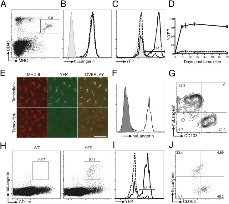
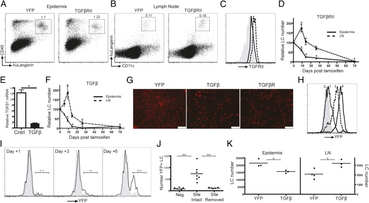
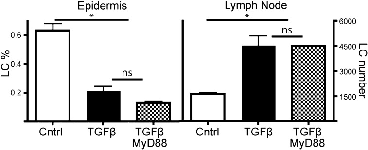
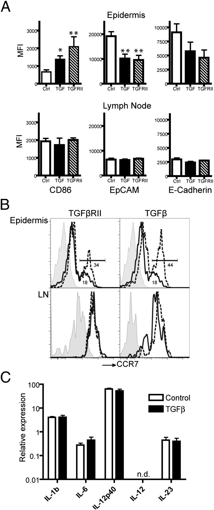
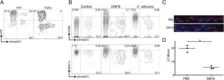
Langerhans cells (LCs) are skin-resident dendritic cells (DC) located in the epidermis that migrate to skin-draining lymph nodes during the steady state and in response to inflammatory stimuli. TGF-β1 is a critical immune regulator that is highly expressed by LCs. The ability to test the functional importance of LC-derived TGF-β1 is complicated by the requirement of TGF-β1 for LC development and by the absence of LCs in mice with an LC-specific ablation of TGF-β1 or its receptor. To overcome these problems, we have engineered transgenic huLangerin-CreER(T2) mice that allow for inducible LC-specific excision. Highly efficient and LC-specific expression was confirmed in mice bred onto a YFP Cre reporter strain. We next generated huLangerin-CreER(T2) × TGF-βRII(fl) and huLangerin-CreER(T2) × TGF-β1(fl) mice. Excision of the TGFβRII or TGFβ1 genes induced mass migration of LCs to the regional lymph node. Expression of costimulatory markers and inflammatory cytokines was unaffected, consistent with homeostatic migration. In addition, levels of p-SMAD2/3 were decreased in LCs from wild-type mice before inflammation-induced migration. We conclude that TGF-β1 acts directly on LCs in an autocrine/paracrine manner to inhibit steady-state and inflammation-induced migration. This is a readily targetable pathway with potential therapeutic implications for skin disease.


Conflict of interest statement

The authors declare no conflict of interest.

Figures

Fig. 1.
Fig. 1.
Specificity and efficiency of huLangerin-CreERT2. (A) Flow cytometry of epidermal single-cell suspension from huLangerin-CreERT2 YFP mice stained with MHC II and CD45 to identify LCs. (B and C) Expression of (B) huLangerin and (C) YFP in LCs from tamoxifen-treated wild-type (shaded), untreated YFP (dashed line), and tamoxifen-treated YFP mice (solid line). (D) Percent of LCs expressing YFP over time in treated (solid line) and untreated (dashed line) YFP mice. (E) Immunofluorescence of epidermal whole mounts stained for YFP (green) and MHC-II (red) from untreated and tamoxifen-treated YFP mice. (Scale bar, 30 μm.) (F) Expression of huLangerin on singlets gated on MHC-II and muLangerin from the dermis of YFP (solid line) or littermate control (shaded) mice. (G) Expression of huLangerin and CD103 from YFP mice gated as in F. (H) Singlets gated cells from skin-draining LNs from wild-type (Left) and YFP (Right) mice stained for CD11c and huLangerin. (I) Expression of YFP in huLangerin+ cells as gated in H from untreated YFP (dashed line) and tamoxifen-treated YFP mice (solid line). (J) CD103 and huLangerin expression on CD11c+ muLangerin+ cells isolated from skin-draining LNs of YFP mice. All data are representative of at least three independent experiments.
Fig. 2.
Fig. 2.
LC-specific inducible excision of TGF-βRII or TGF-β1 results in LCs migration. Flow cytometry of (A) epidermis and (B) LNs from YFP (Left) and TGF-βRII (Right) mice on day +9 after tamoxifen injection. (C) Expression of TGF-βRII on LCs gated as in B, isolated from LNs of tamoxifen-treated (solid line) or untreated (dashed line) TGF-βRII mice. The shaded line represents isotype control. (D) Relative number of LCs in the epidermis (solid line) or skin-draining LNs (dashed line) of TGF-βRII mice at the indicated number of days after tamoxifen treatment, as a percentage compared with control YFP mice. (E) Relative expression of TGF-β1 mRNA in LCs isolated from untreated (Cntl) or tamoxifen-treated TGF-β mice. (F) Relative number of LCs in the epidermis (solid line) or skin-draining LNs (dashed line) from tamoxifen-treated TGF-β1 mice, as in D. (G) Immunofluorescence of epidermal whole mounts stained for MHC-II (red) isolated from YFP, TGF-β, and TGF-βRII mice on day +17 after tamoxifen treatment. (Original magnification: 100×; scale bar, 100 μm.) (H) YFP expression in LCs isolated from the epidermis of YFP mice after a single topical application of tamoxifen. Numbers indicate the number of days after tamoxifen application. (I) Percentage of YFP+ LCs isolated from the cervical LNs of YFP mice at the indicated day after a single application of topical tamoxifen. (J) Ears of YFP mice were painted with vehicle (Neg) or tamoxifen and either left intact (Site intact) or surgically excised after 18 h (Site removed). The number of YFP+ LCs isolated from cervical LNs on day +6 is shown. Each symbol represents an individual animal. (K) Number of epidermal YFP+ LCs per ear (Left) and ipsilateral cervical LNs (Right) from YFP and TGF-β mice on day 9 after topical tamoxifen application. Each symbol represents an individual animal. All data are representative of at least three independent experiments with cohorts of three to six mice except for C, E, and H, which used three individual mice. *P < 0.05; **P < 0.01.
Fig. 3.
Fig. 3.
LC migration in response to loss of TGF-β1 is MyD88 independent. Percentage of LCs found in the epidermis (Left) and number of LCs found in skin-draining LNs (Right) of YFP (Cntl), TGF-β, and TGF-β/MyD88 mice on day +9 after i.p. administration of tamoxifen. Data are representative of three independent experiments with cohorts of three to five mice. ns, not significant. *P < 0.05.
Fig. 4.
Fig. 4.
TGF-β1–deficient LCs maintain steady-state phenotype. (A) Mean fluorescence intensity of CD86, EpCAM, and E-cadherin staining for LCs isolated from the epidermis and gated as CD45+ MHC-II+ (Upper) or skin-draining lymph node gates as CD11c+, huLangerin+ (Lower) of TGF-β, TGF-βRII and control mice on day +9 after tamoxifen treatment. Histograms from representative mice are shown in Fig. S4. Data are representative of at least three independent experiments with groups of four to five mice each. (B) Expression of CCR7 on LCs isolated from epidermis (Upper) or LNs (Lower) on day +9 after tamoxifen treatment. Cells from YFP mice are represented by solid lines, whereas cells from TGF-βRII (Left) or TGF-β (Right) are shown as broken lines. Percentage of CCR7 cells is indicated. Shaded lines are isotype staining. (C) Relative expression of IL-1β, IL-6, IL-12β (IL-12p40), IL-12α (IL-12p35), and IL-23 is shown as assessed by quantitative PCR of mRNA isolated from LCs in the skin-draining LNs of control and TGF-β mice. Data are from two independent experiments. n.d., not detected. *P < 0.05, **P < 0.01.
Fig. 5.
Fig. 5.
TGF-β signaling is interrupted during inflammation. (A) Expression of pSMAD2/3 and YFP expression in LCs (CD45+, MHC-II+), in cells isolated from unmanipulated YFP and TGF-β mice on day +9 after tamoxifen treatment. (B) Expression of CD86 (Upper) and MHC II (Lower) vs. pSMAD2/3 is shown in LCs isolated from the epidermis of wild-type mice 18 h after application of 0.5% DNFB or infection with C. albicans (24, 26). Data represent two independent experiments with groups of three mice each. (C) Wild-type mice were treated with the TGF-βRI kinase inhibitor SM16 for 47 d. Immunofluorescence of transverse sections of ear skin from PBS- and SM16-treated mice is shown. (D) Number of LCs per millimeter of epidermis. Each symbol represents an individual animal. *P < 0.05.

References

    1. Romani N, Clausen BE, Stoitzner P. Langerhans cells and more: Langerin-expressing dendritic cell subsets in the skin. Immunol Rev. 2010;234:120–141. - PMC - PubMed
    1. Hemmi H, et al. Skin antigens in the steady state are trafficked to regional lymph nodes by transforming growth factor-beta1-dependent cells. Int Immunol. 2001;13:695–704. - PubMed
    1. Merad M, Ginhoux F, Collin M. Origin, homeostasis and function of Langerhans cells and other langerin-expressing dendritic cells. Nat Rev Immunol. 2008;8:935–947. - PubMed
    1. Chorro L, et al. Langerhans cell (LC) proliferation mediates neonatal development, homeostasis, and inflammation-associated expansion of the epidermal LC network. J Exp Med. 2009;206:3089–3100. - PMC - PubMed
    1. Merad M, et al. Langerhans cells renew in the skin throughout life under steady-state conditions. Nat Immunol. 2002;3:1135–1141. - PMC - PubMed

Publication types

Substances

LinkOut - more resources