

Disseminated penicilliosis in a Korean human immunodeficiency virus infected patient from Laos
- PMID: 22690104
- PMCID: PMC3369459
- DOI: 10.3346/jkms.2012.27.6.697
Disseminated penicilliosis in a Korean human immunodeficiency virus infected patient from Laos
Abstract
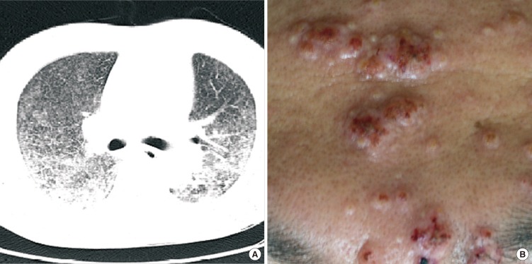
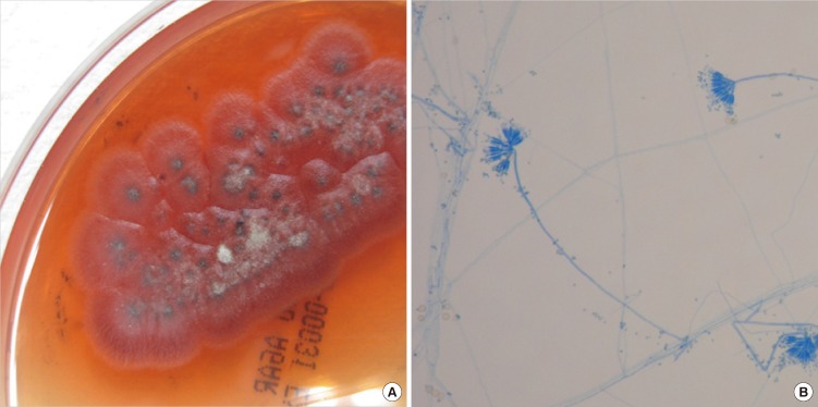
Penicillium marneffei may cause life-threatening systemic fungal infection in immune-compromised patients and it is endemic in Southeast Asia. A 39-yr-old HIV-infected male, living in Laos, presented with fever, cough, and facial vesiculopapular lesions, which had been apparent for two weeks. CT scans showed bilateral micronodules on both lungs; Pneumocystis jirovecii was identified by bronchoscopic biopsy. Despite trimethoprim-sulfamethoxazole and anti-tuberculosis medications, the lung lesions progressed and the facial lesions revealed central umbilications. Biopsy of the skin lesions confirmed disseminated penicilliosis, with the culture showing P. marneffei hyphae and spores. The P. marneffei was identified by rRNA PCR. A review of the bronchoscopic biopsy indicated penicilliosis. The patient completely recovered after being prescribed amphotericin-B and receiving antiretroviral therapy. This is the first case of penicilliosis in a Korean HIV-infected patient. It is necessary to consider P. marneffei when immunocompromised patients, with a history of visits to endemic areas, reveal respiratory disease.
Keywords: Disseminated Infection; HIV/AIDS; Korean; Penicillium marneffei.
Figures
References
-
- Capponi M, Sureau P, Segretain G. Penicilliosis de Rhizomys sinensis. Bull Sco Pathol Exot Filiales. 1956;49:418–421. - PubMed
-
- DiSalvo AF, Ficking AM, Ajello L. Infection caused by Penicillium marneffei: description of first natural infection in man. Am J Clin Pathol. 1973;60:259–263. - PubMed
-
- Piehl MR, Kaplan RL, Haber MH. Disseminated penicilliosis in a patient with acquired immunodeficiency syndrome. Arch Pathol Lab Med. 1988;112:1262–1264. - PubMed
-
- Supparatpinyo K, Khamwan C, Baosoung V, Nelson KE, Sirisanthana T. Disseminated Penicillium marneffei infection in southeast Asia. Lancet. 1994;344:110–113. - PubMed
-
- Yap FB, Thevarajah S, Asmah J. Penicillium marneffei infection in an African man. Dermatol Online J. 2010;16:2. - PubMed
Publication types
MeSH terms
Substances
LinkOut - more resources
Full Text Sources
Medical
