. 2012:18:1402-10.
Epub 2012 Jun 1.

WW Domain Containing Transcription Regulator regulates human conjunctiva epithelial cell proliferation via inhibiting TGFβ signaling pathway [corrected]

Affiliations

WW Domain Containing Transcription Regulator regulates human conjunctiva epithelial cell proliferation via inhibiting TGFβ signaling pathway [corrected]

Xiao Wei Tan et al. Mol Vis. 2012.

Erratum in

  • Mol Vis. 2014;20:284

Abstract

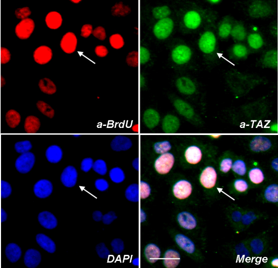
Purpose: To investigate the role of Tafazzin (TAZ) protein in regulating the proliferation of normal human conjunctiva epithelial cells and epithelial cells from pterygium tissue.

Methods: Conjunctiva epithelial cells were cultured in keratinocytes growth medium and treated with transformation growth factor β (TGFβ) to analyze the expression and translocation of TAZ protein by immunostaining and BrdU analysis. Immortalized conjunctiva epithelial cells (NHC) were treated with TGFβ, targeting siRNA, TGFβ receptor antibody or TGFβ receptor inhibitor, to study the involvement of TAZ and TGFβ signaling pathway in conjunctiva cell proliferation by cell adhesion assay. Conjunctiva tissues from a normal human eye and an eye with pterygium disease were collected for histological analyses and western blot to evaluate the TAZ protein expression in vivo.

Results: TAZ expression was upregulated in mitotic conjunctiva epithelial cells, proliferating conjunctiva epithelial cells, TGFβ treated conjunctiva epithelial cells and human pterygium epithelium. TAZ siRNA induced less conjunctiva epithelial cell growth. Moreover, TGFβ receptor antibody and TGFβ receptor inhibitor rescued this anti-proliferative effect of TAZ siRNA.

Conclusions: TAZ is involved in human conjunctiva epithelial cells proliferation via regulating TGFβ signaling pathway.


Figures

Figure 1
Figure 1
TAZ protein expression is upregulated in mitotic cells. A: Conjunctiva epithelial cells were stained with anti-TAZ antibody. TAZ: red; DAPI: blue. Higher magnification image is shown in lower row. Immunofluorescence signal of TAZ protein is significantly higher in a dividing cell (white arrow) compared with that of a neighbor cell (white star). Scale bar is 20 µm. B: Relative quantitative red fluorescence intensity of total 30 dividing cells and 90 neighbor cells were measured. Data were represented as mean±se ** p<0.05, one-way ANOVA (ANOVA).
Figure 2
Figure 2
TAZ protein expression is upregulated in mitotic cells at various cell cycle stage. A and B: Conjunctiva epithelial cells were stained by TAZ (red) after treating with hydrochloride for 15 min (A) or 40 min (B). Counterstaining with DAPI (blue) was performed as an indicator of cells at different cell cycle stages. White arrows indicate the cells which were undergoing cell division. Scale bar is 20 µm. C: TAZ siRNA induced less 10–4-cyclin E promoter transcription activities in NHC cell lines in a dosage dependent manner. Data have been represented as mean±se from three replicates.
Figure 3
Figure 3
TAZ protein clusters at the nucleus of proliferating epithelial cells. Conjunctiva epithelial cells were labeled for BrdU (red) and TAZ (green), DAPI was applied as nucleus counter staining dye (blue). White arrows indicate a BrdU positive epithelial cell also stained by high intensity of TAZ protein. Scale bar is 20 µm.
Figure 4
Figure 4
TAZ is responsive to TGFβ signal in conjunctiva epithelial cells. A: Fluorescent signals of TAZ protein (green) and smad4 (red) were upregulated after TGFβ treatment. Scale bar is 20 µm. B: Relative quantitation of the green fluorescent intensity of TAZ protein. Data were represented as mean±se, * p<0.01, ANOVA. C: Cell lysate of the epithelial cells with or without TGFβ treatment were blotted by anti-TAZ. Expression of TAZ protein was upregulated in TGFβ treated cells.
Figure 5
Figure 5
TAZ siRNA induced a depletion of TAZ protein in NHC keratinocytes. A: Anti-TAZ blotting of the NHC cells which were treated by 15 PM TAZ siRNA or mock siRNA. B: Pictures show representative fields of cells treated by mock or TAZ targeting siRNA. NHC cells were stained by commassie brilliant blue. Scale bar is 10 um.
Figure 6
Figure 6
TGFβ receptor antibody and TGFβ receptor inhibitor rescued the effect of TAZ siRNA on cell growth. A: NHC cells were treated with TGFβ receptor type I antibody together with mock siRNA or TAZ siRNA. Relative cell density was analyzed from three replicates. a: a significant reduction of cell density after TAZ siRNA treatment. “b”: the cell growth inhibition was rescued by TGFβ receptor I antibody treatment. B: NHC cells were treated with TGFβ receptor inhibitor (SB431542) together with mock siRNA or TAZ siRNA. Relative cell density was analyzed from three replicates. “a’”: a significant reduction of cell density after TAZ siRNA treatment. “b’”: cell growth inhibition was rescued by SB431542. Data were represented as mean±se (p<0.05).
Figure 7
Figure 7
Location and expression of TAZ proteins in normal or pterygium conjunctiva epithelium. A: Total tissue lysate from normal or human conjunctiva tissue with pterygium disease were blotted with anti-TAZ (A). Relative intensity of the blots was measured from three replicates (B). C: Conjunctiva tissue was stained by anti-TAZ (red) and DAPI (blue). Merged images show that the TAZ protein is expressed at the epithelial layer of conjunctiva tissue. Scale bar is 10 µm.

References

    1. Golu T, Mogoanta L, Streba CT, Pirici DN, Malaescu D, Mateescu GO, Mutiu G. Pterygium: histological and immunohistochemical aspects. Rom J Morphol Embryol. 2011;52:153–8. - PubMed
    1. Kase S, Takahashi S, Sato I, Nakanishi K, Yoshida K, Ohno S. Expression of p27(KIP1) and cyclin D1, and cell proliferation in human pterygium. Br J Ophthalmol. 2007;91:958–61. - PMC - PubMed
    1. Shi Y, Massague J. Mechanisms of TGF-beta signaling from cell membrane to the nucleus. Cell. 2003;113:685–700. - PubMed
    1. Siegel PM, Massague J. Cytostatic and apoptotic actions of TGF-beta in homeostasis and cancer. Nat Rev Cancer. 2003;3:807–21. - PubMed
    1. Ten Dijke P, Goumans MJ, Itoh F, Itoh S. Regulation of cell proliferation by Smad proteins. J Cell Physiol. 2002;191:1–16. - PubMed

MeSH terms

LinkOut - more resources