Case Reports
. 2012 Jan;6(1):1-8.
doi: 10.3941/jrcr.v6i1.863. Epub 2012 Jan 1.

A case of Rosai-Dorfman disease in a pediatric patient with cardiac involvement

Affiliations
Case Reports

A case of Rosai-Dorfman disease in a pediatric patient with cardiac involvement

Leonard Yontz et al. J Radiol Case Rep. 2012 Jan.

Abstract

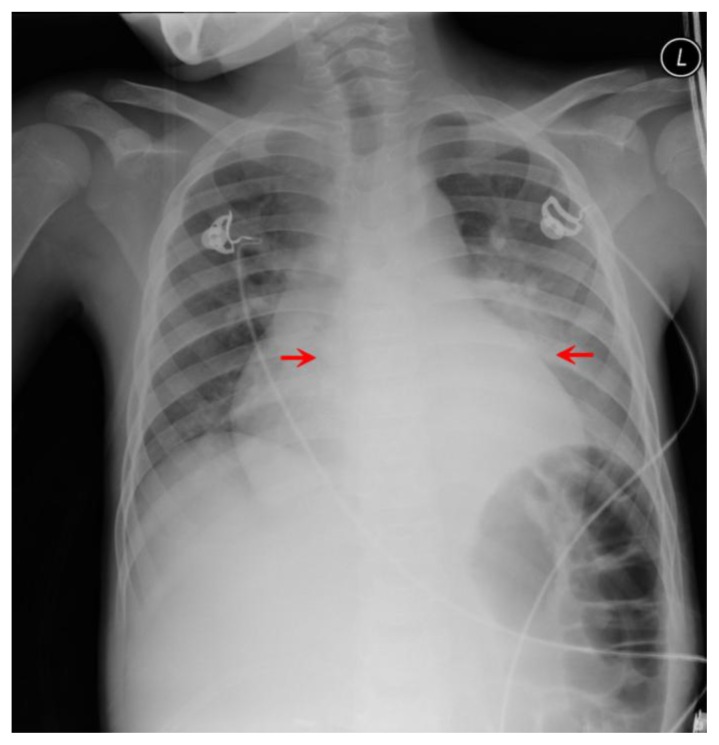
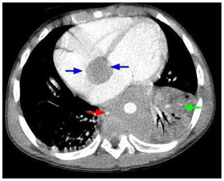
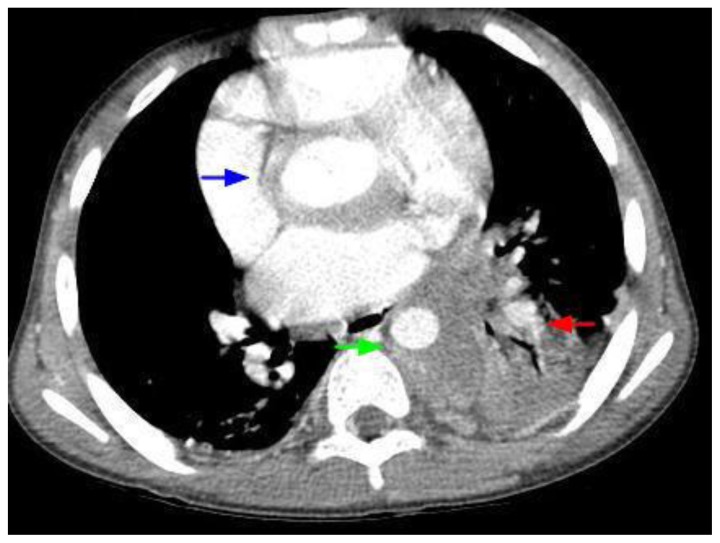
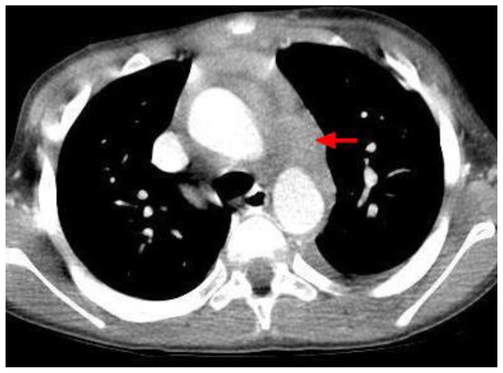
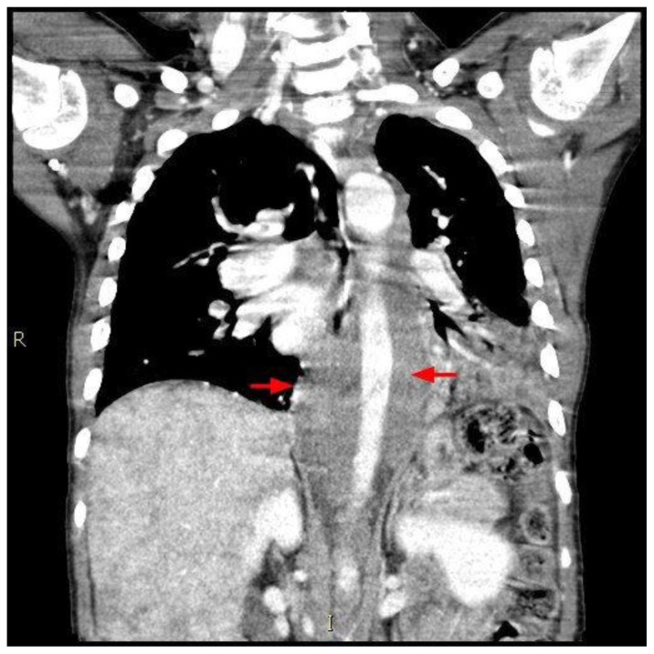
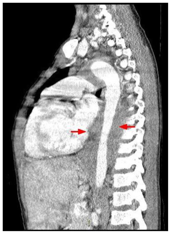
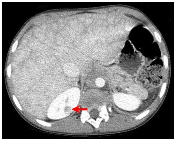
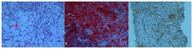
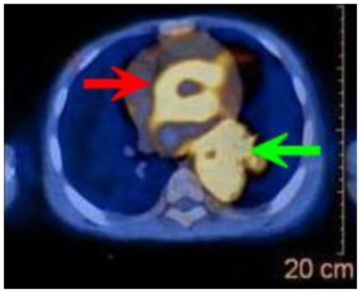
Rosai-Dorfman disease (RDD) involves abnormal proliferations of oddly behaving histocytes that are not derived from the Langerhan's Cell linage. These collections tend to occur within lymph nodes, with occasional extra nodal presentation. While RDD is a rare entity itself, extra nodal cases are even more so, with even fewer reporting cardiac involvement, and previously only in adults. This report describes the disease in a pediatric patient who had the unique feature of an extra nodal cardiac mass. The patient, who was known to have sickle cell disease, was initially erroneously thought to have acute chest syndrome. Sudden changes in the patient's status, including development of 3rd degree heart block, demanded investigation with additional imaging. Chest CT revealed a mass arising from the cardiac interatrial septum and encircling the entire thoracic aorta. Imaging features of Rosai-Dorfman disease are nonspecific, complicating the diagnosis. We present this case with discussion of this extremely uncommon entity. We describe the diagnostic methods, the differential diagnosis, and the treatment options.

Keywords: Heart; Rosai-Dorfman Disease.


Figures

Figure 1
Figure 1
12-year-old male with Rosai-Dorfman disease. Frontal radiograph of the chest reveals a magnified cardiac silhouette and retrocardiac opacity (red arrows).
Figure 2
Figure 2
12-year-old male with Rosai-Dorfman disease. Axial chest CT obtained following the intravenous administration of 100 ml of Omnipaque 350. The CT setting was 100 kVp with modulated mAs. The image reveals a cardiac mass originating from the interatrial septum and abutting the atrioventricular groove (blue arrows). There is soft tissue density encasing the descending aorta (red arrow). There is left lower lobe atelectasis (green arrow) due to compression of the left lower lobe bronchus by the mass.
Figure 3
Figure 3
12-year-old male with Rosai-Dorfman disease. Axial chest CT obtained following the intravenous administration of 100 ml of Omnipaque 350. The CT setting was 100 kVp with modulated mAs. The image reveals a mass encasing the aortic root (blue arrow). There is left lower lobe atelectasis (red arrow) secondary to compression of the left lower lobe bronchus. The mass is noted to encircle the descending aorta (green arrow) and compress the esophagus.
Figure 4
Figure 4
12-year-old male with Rosai-Dorfman disease. Axial chest CT obtained following the intravenous administration of 100 ml of Omnipaque 350. The CT setting was 100 kVp with modulated mAs. The image reveals a mass encircling the aortic arch (red arrow) and the ascending and descending aorta.
Figure 5
Figure 5
12-year-old male with Rosai-Dorfman disease. Coronal chest CT obtained following the intravenous administration of 100 ml of Omnipaque 350. The CT setting was 100 kVp with modulated mAs. The image reveals a soft tissue mass encasing the descending aorta (red arrows). The mass extend to the level of the upper abdomen. Note the mottled appearance of the liver.
Figure 6
Figure 6
12-year-old male with Rosai-Dorfman disease. Sagittal chest CT obtained following the intravenous administration of 100 ml of Omnipaque 350. The CT setting was 100 kVp with modulated mAs. The image reveals a soft tissue mass encasing the descending aorta (red arrows). The mass extend to the level of the upper abdomen. Note the mottled appearance of the liver.
Figure 7
Figure 7
12-year-old male with Rosai-Dorfman disease. Axial chest CT obtained following the intravenous administration of 100 ml of Omnipaque 350. The CT setting was 100 kVp with modulated mAs. The image reveals a complex cyst with enhancing septa in the upper pole of the right kidney (red arrow). Note the mottled appearance of the liver.
Figure 8
Figure 8
12-year-old male with Rosai-Dorfman disease. (a) Section demonstrating large bland histiocytic cells with pale to foamy cytoplasm displaying emperipolesis (red arrow), admixed with mature lymphocytes, plasma cells and occasional polymorphs. Mild delicate myofibroblastic proliferation is noted in a reactive pattern (H&E × 100). (b) Histiocytic cells are intensely immunoreactive for S-100 protein (S-100 × 200). (c) Immunostain for smooth muscle actin highlights the delicate myofibroblastic proliferation in a reactive pattern with relatively scant collagen deposition (SMA × 100).
Figure 9
Figure 9
12-year-old male with Rosai-Dorfman disease. Axial view of a PET/CT obtained following the intravenous administration of 8.7 mCi of F-18-FDG. The enhanced CT portion of the study was obtained following the intravenous administration of 100 ml of Omnipaque 350. The CT setting was 100 kVp with modulated mAs. There is intense hypermetabolism in the mass that surrounds the ascending and the descending aorta. The maximal SUV measured in the mass was 9.8, significantly higher than the blood pool activity.

References

    1. McClain K, Natkunam Y, Swerdlow S. Atypical Cellular Disorders Hematology the American Society of Hematology Education Program Book. 2004:283–96. - PubMed
    1. Rosia J, Dorfman RF. Sinus Histiocytosis with Massive Lymphadenophathy. A newly recognized benign clinicopathological entity. Archives of Pathology. 1969;87(1):63–70. - PubMed
    1. Pulsoni A, et al. Treatment of Sinus Histiocytosis with Massive Lymphadenopathy (Rosai-Dorfman Disease): Report of a case and Literature Review. American Journal of Hematology. 2002;69:67–71. - PubMed
    1. La Barge D, III, Salzman K, Harnsberger H, et al. Sinus Histiocytosis with Massive Lymphadenopathy (Rosai-Dorfman Disease): Imaging Manifestations in the Head and Neck. American Journal of Roentgenology. 2008;191:W299–306. - PubMed
    1. Molina-Garrido MJ. Guillén-Ponce C, Extranodal Rosai-Dorfman Disease with Cutaneous and Periodontal Involvement: A Rare Presentation. Case Reports Oncology. 2011;4:96–100. - PMC - PubMed

Publication types

LinkOut - more resources