Case Reports
. 2012 Jan;6(1):31-7.
doi: 10.3941/jrcr.v6i1.889. Epub 2012 Jan 1.

3-D printout of a DICOM file to aid surgical planning in a 6 year old patient with a large scapular osteochondroma complicating congenital diaphyseal aclasia

Affiliations
Case Reports

3-D printout of a DICOM file to aid surgical planning in a 6 year old patient with a large scapular osteochondroma complicating congenital diaphyseal aclasia

Matthew D Tam et al. J Radiol Case Rep. 2012 Jan.

Abstract

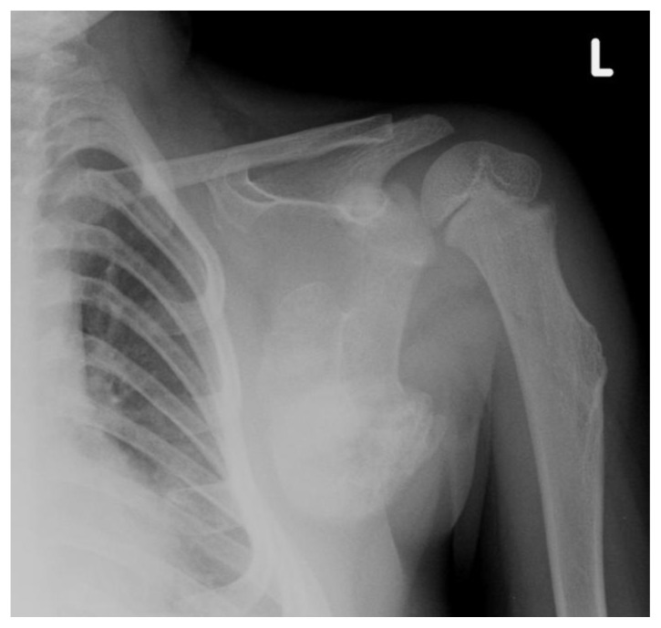
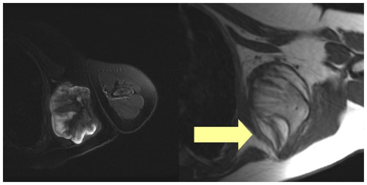
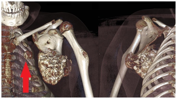
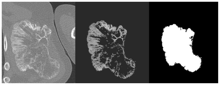
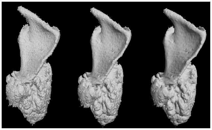
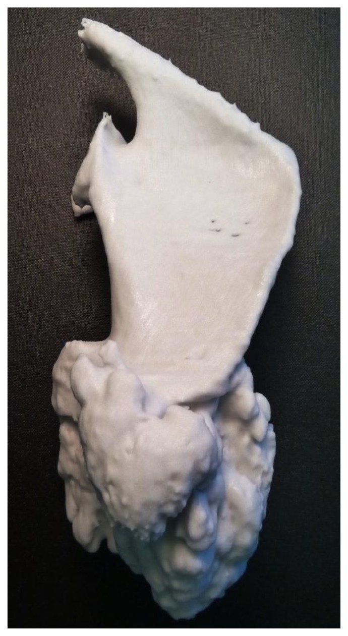
A 6 year old girl presented with a large osteochondroma arising from the scapula. Radiographs, CT and MRI were performed to assess the lesion and to determine whether the lesion could be safely resected. A model of the scapula was created by post-processing the DICOM file and using a 3-D printer. The CT images were segmented and the images were then manually edited using a graphics tablet, and then an STL-file was generated and a 3-D plaster model printed. The model allowed better anatomical understanding of the lesion and helped plan surgical management.

Keywords: 3D modelling; 3D printing; DICOM; anatomy; diaphyseal aclasia; image processing; model; rapid prototyping; scapular osteochondroma; segmentation; surgical planning.


Figures

Figure 1
Figure 1
A 6 year-old girl with a large scapular osteochondroma complicating congenital diaphyseal aclasia. Plain radiograph of the left shoulder girdle demonstrates osteochondromata arising from the scapula and also humeral shaft.
Figure 2
Figure 2
A 6 year-old female with a large scapular osteochondroma complicating congenital diaphyseal aclasia. On the left, axial T2-fat saturated images demonstrate the hyperintense cartilage cap (Protocol: 1.5 T, TE 830888, TR 3960, slice thickness 5 mm, slice spacing 7 mm, matrix 0/320/256/0, echo train 16). On the right, the axial T1 weighted images with an arrow demonstrating the serratus anterior (Protocol: 1.5 T, TE 14, TR 420, slice thickness 5 mm, slice spacing 7 mm, matrix 0/256/224/0, echo train 0).
Figure 3
Figure 3
A 6 year-old girl with a large scapular osteochondroma complicating congenital diaphyseal aclasia. Volume rendered images from the non-contrast CT demonstrate anterior and posterior views the lesion arising from the inferior angle of the scapula. Note also another osteochondroma arising from the 2nd anterior rib (arrow). (Protocol: 128 slice non-contrast CT, slice thickness 1mm, kVp 120, mAs 29).
Figure 4
Figure 4
A 6 year-old girl with a large scapular osteochondroma complicating congenital diaphyseal aclasia. Stages of image-processing (a) DICOM CT image (b) threshold segmentation (c) final conversion with manual editing into a black and white STL file.
Figure 5
Figure 5
A 6 year-old girl with a large scapular osteochondroma complicating congenital diaphyseal aclasia. Pre-print 3-D computer modelling with increasing number of Marching Cube algorithms applied from left to right (posterior views).
Figure 6
Figure 6
A 6 year-old girl with a large scapular osteochondroma complicating congenital diaphyseal aclasia. Photograph of the plaster model (posterior view).

References

    1. Graham RN, Perriss RW, Scarsbrook AF. DICOM demystified: a review of digital file formats and their use in radiological practice. Clin Radiol. 2005;60(11):1133–40. - PubMed
    1. Rosset A, Spadola L, Ratib O. OsiriX: an open-source software for navigating in multidimensional DICOM images. J Digit Imaging. 2004;17(3):205–216. - PMC - PubMed
    1. Lorensen W, Cline H. Marching cubes: A high resolution 3D surface construction algorithm. SIGGRAPH Comput Graph. 1987;21(4):163–169.
    1. Rengier F, Mehndiratta A, von Tengg-Kobligk H, Zechmann CM, Unterhinninghofen R, Kauczor HU, Giesel FL. 3D printing based on imaging data: review of medical applications. Int J Comput Assist Radiol Surg. 2010;5(4):335–41. - PubMed
    1. Jacobs S, Grunert R, Mohr F, Falk V. 3D-Imaging of cardiac structures using 3D heart models for planning in heart surgery: a preliminary study. Interactive CardioVascular and Thoracic Surgery. 2008;7:6–9. - PubMed

Publication types

MeSH terms