Review
. 2012 Mar;6(3):1-8.
doi: 10.3941/jrcr.v6i3.745. Epub 2012 Mar 1.

NOMID: the radiographic and MRI features and review of literature

Affiliations
Review

NOMID: the radiographic and MRI features and review of literature

Faizah Mohd Zaki et al. J Radiol Case Rep. 2012 Mar.

Abstract

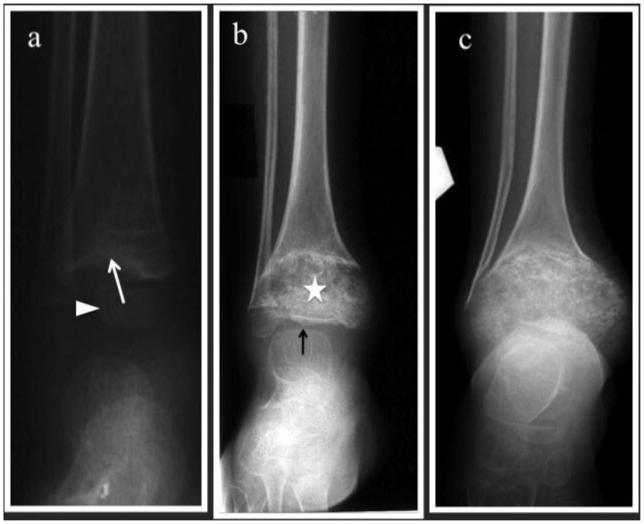
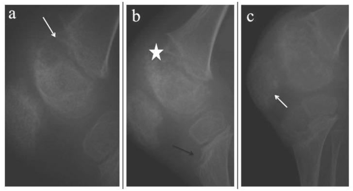
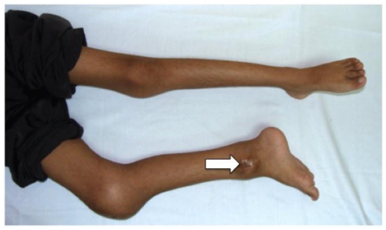
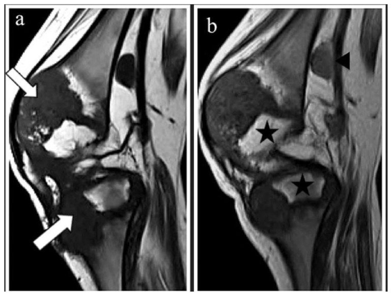
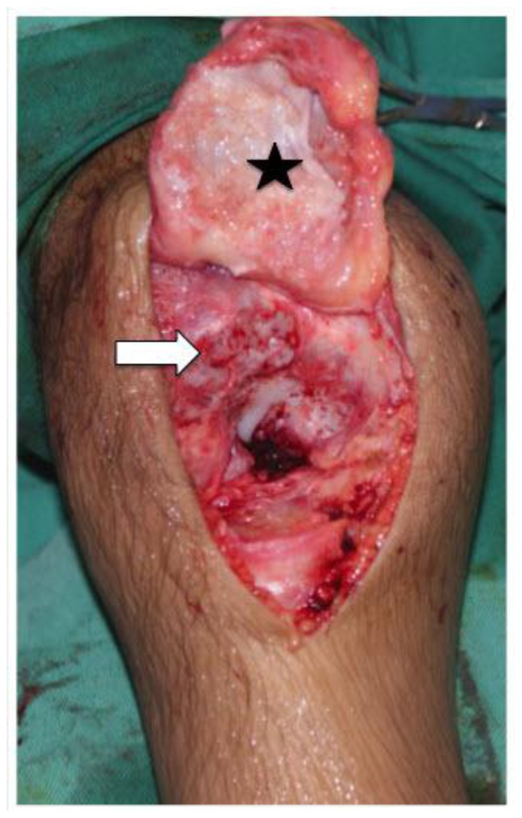
Neonatal onset multisystem inflammatory disease (NOMID) is a rare autoinflammatory disorder, which manifests early in infancy. We describe a case of a 10-year-old boy who has been unwell since infancy. He presented with urticarial rash, intermittent fever and hepatosplenomegaly followed by progressive arthropathy. His joint symptoms started at two years of age, which progressively involved multiple joints, resulting in bone and joint deformities. A series of joint radiographs demonstrated bizarre enlarging physeal mass with heterogenous calcification. Magnetic resonance imaging (MRI) of the involved right ankle and knee showed characteristic thickened and calcified physeal lesions, which enhanced post-gadolinium. This debilitating disease is also known to involve the central nervous system and eyes. This case report aims to highlight the conventional radiographic and magnetic resonance imaging (MRI) findings of this physeal abnormality in NOMID syndrome.

Keywords: CINCA; MRI; NOMID; Neonate; radiograph.


Figures

Figure 1
Figure 1
10-year-old boy with Neonatal Onset Multisystem Inflammatory Disease (NOMID). A series of antero-posterior radiographs of the right ankle which were done at 2, 6 and 9 years of age (a-c) respectively. (a) Eccentrically located uncalcified distal tibial physeal mass indenting the metaphysis. The metaphyseal margin appears sclerotic (white arrow). Epiphysis is normal and labeled with white arrowhead. (b) Mass-like physis with heterogenous calcification (white star). The metaphysis shows cupping and flaring. The residual epiphysis is flattened and compressed (white arrow) (c) Progressive enlargement of the calcified physis. The epiphysis is no longer seen and there is progressive cupping of the metaphysis to accommodate the enlarged physis.
Figure 2
Figure 2
10-year-old boy with Neonatal Onset Multisystem Inflammatory Disease (NOMID). Series of lateral right knee radiographs done at 5, 9 and 10 years of age respectively (a-c) (a) At 5 years old, there is cupping of the metaphysis (white arrow) by uncalcified physeal mass with associated cupping of the distal epiphysis. (b-c) By the age of 9 – 10 years old, the enlarged physis has calcified causing mass-like heterogenous ossification (white star), which is indistinct from the adjacent epiphysis. The proximal tibial metaphysis begins to show eccentric anterior cupping caused by uncalcified physeal mass (black arrow). Patella is enlarged with irregular outline (white arrow)
Figure 3
Figure 3
10-year-old boy with Neonatal Onset Multisystem Inflammatory Disease (NOMID). Picture of the child’s lower limbs that shows swelling of the right knee and ankle joints. There is also flexion deformity of the right knee joint. An ulcer is seen at the enlarged right ankle due to a previous biopsy (white arrow).
Figure 4
Figure 4
10-year-old boy with Neonatal Onset Multisystem Inflammatory Disease (NOMID). Sagittal MRI of the right knee done at the age of 10 years old: (a) T1WI-pre gadolinium (1.5 Tesla, TR/TE=400msec/8msec, Slice Thickness=3mm); and (b) post gadolinium (1.5 Tesla, TR/TE=400msec/8msec, Slice Thickness=3mm; Omniscan®, 0.2mls/kg). (a) The eccentrically enlarged physis of the distal femur and proximal tibia are predominantly hypointense due to the presence of calcification (white arrow) (b) There is minimal enhancement post contrast. Note the compressed epiphysis of the distal femur and proximal tibia (black stars). An enlarged popliteal node is also noted, measuring 0.8cmx1.0cmx1.5cm (anteroposterior × width × craniocaudal)(black arrowhead). The patellofemoral joint space is obliterated.
Figure 5
Figure 5
10-year-old boy with Neonatal Onset Multisystem Inflammatory Disease (NOMID). Intraoperative photograph of the right knee shows the denuded cartilage of the patella & distal femur. The patellofemoral joint was ankylosed. The patella ligament was released (black star) & the ankylosed joint osteotomized to separate the patella from the femur (white arrow).

References

    1. Prieur AM, Griscelli C, Lampert F, Trunkenbrodt H, Guggenheim MA, Lovell DJ, et al. A Chronic, Infantile, Neurological, Cutaneous and Articular (CINCA) Syndrome. A specific entity analysed in 30 patients. Scand J Rheumatology. 1987;(Suppl 66):57–68. - PubMed
    1. Prieur AM, Griscelli C. Arthropathy with rash, chronic meningitis, eye lesions, and mental retardation. The Journal of Pediatrics. 1981;99(1):79–83. - PubMed
    1. Kaufman RA, Lovell DJ. Infantile-onset Multisystem Inflammatory Disease: Radiologic findings. Radiology. 1986;160:741–746. - PubMed
    1. Hill SC, Namde M, Dwyer A, Poznanski A, Canna S, Goldbach-Mansky R. Arthropathy of neonatal onset multisystem inflammatory disease (NOMID/CINCA) Pediatric Radiology. 2007;37:145–152. - PubMed
    1. Miyamae T, Inaba Y, Nishimura G, Kikuchi M, Kishi T, Hara R, et al. Effect of anakinra on arthropathy in CINCA/NOMID syndrome. Pediatric Rheumatology. 2001;8:9. Available from http://www.ped-rheum.com/content/8/1/9.pdf. - PMC - PubMed

LinkOut - more resources