. 2012 Jul 6;3(1):11.
doi: 10.1186/2041-9139-3-11.

Mechanisms of stomatal development: an evolutionary view

Affiliations

Mechanisms of stomatal development: an evolutionary view

Anne Vatén et al. Evodevo. .

Erratum in

Abstract

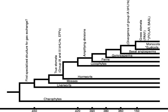
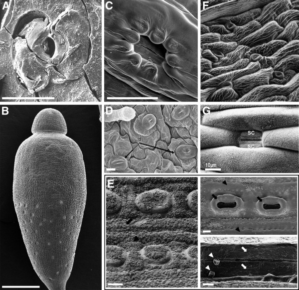
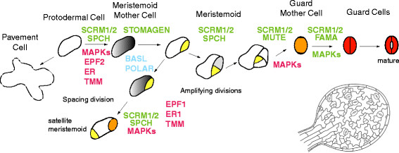
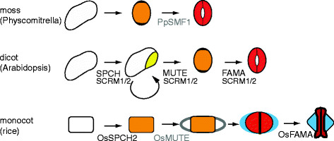
Plant development has a significant postembryonic phase that is guided heavily by interactions between the plant and the outside environment. This interplay is particularly evident in the development, pattern and function of stomata, epidermal pores on the aerial surfaces of land plants. Stomata have been found in fossils dating from more than 400 million years ago. Strikingly, the morphology of the individual stomatal complex is largely unchanged, but the sizes, numbers and arrangements of stomata and their surrounding cells have diversified tremendously. In many plants, stomata arise from specialized and transient stem-cell like compartments on the leaf. Studies in the flowering plant Arabidopsis thaliana have established a basic molecular framework for the acquisition of cell fate and generation of cell polarity in these compartments, as well as describing some of the key signals and receptors required to produce stomata in organized patterns and in environmentally optimized numbers. Here we present parallel analyses of stomatal developmental pathways at morphological and molecular levels and describe the innovations made by particular clades of plants.


Figures

Figure 1
Figure 1
Divergence of major land plant lineages and appearance of stomatal characteristics. Phylogenetic tree of extant land plants indicating positions of major innovations in the evolution of stomata, following Ruszala et al.[5]. Those in brackets indicate predicted appearance of stomatal development regulatory genes. Numbers on the x-axis refer to multiples of millions of years
Figure 2
Figure 2
Representative stomatal complexes and patterns from different species. (A) Scanning electron micrograph (SEM) of Silurian fossil stoma displaying common morphology. Scale bar, 20 μm [3]. (B) SEM of moss Bryum capillare sporangium with stomata visible on the lower half. Scale bar, 600 μm [2]. (C) SEM of moss Bryum capillare sporangium stoma sunken below epidermal cells. Scale bar, 50 μm [2]. (D) SEM of fern Thelypteris ovata var. lindheimeri (sporophyte) leaf with stomata separated by pavement cells. Scale bar, 10 μm; s, stomata [6]. (E) Left panel, field emission SEM of Pinus koraiensis (gymnosperm) stomata arranged in rows on needle surface; granular material is surface wax. Scale bar, 10 μm. Upper right, dewaxed stomata. Scale bar, 10 μm. Lower right, dewaxed guard cells (arrows) within an epistomatal chamber. Scale bar, 2 μm [7]. (F) SEM of dicot Arabidopsis thaliana stomatal pattern in the sepal. (G) SEM of monocot Poa annua stoma, with subsidiary cells (sc) flanking the narrow guard cells. Scale bar, 10 μm [8]
Figure 3
Figure 3
Stomatal development in Arabidopsis . Diagram of major stages in stomatal development with place of action of the subset of regulatory genes discussed in this review noted. Positive regulators are written in green, negative regulators in red and polarity regulators in blue. Not all genes known to regulate stomata are presented. The image of the young leaf in the lower right corner is to represent the dispersed nature of stomatal lineage initiation. Color code: yellow, meristemoid; orange, guard mother cell; red, guard cell; grey, meristemoid mother cell (MMC)
Figure 4
Figure 4
Comparison of the molecular and morphological features of stomatal development in Arabidopsis and representatives of the grasses and mosses for which molecular data exist. Presentation of a simplified stomatal lineage displaying only cell identities (in the same color codes as Figure 3), with the addition of blue to mark the subsidiary cells in monocots. Genetic regulators of the processes are included at their points of action, with black text indicating that there is direct functional evidence supporting the placement and grey text representing inferences from cross-species complementation tests. The curved arrow in the dicot lineage represents the continued asymmetric amplifying divisions made by meristemoids

References

    1. Sirichandra C, Wasilewska A, Vlad F, Valon C, Leung J. The guard cell as a single-cell model towards understanding drought tolerance and abscisic acid action. J Exp Bot. 2009;60:1439–1463. doi: 10.1093/jxb/ern340. - DOI - PubMed
    1. Ligrone R, Duckett JG, Renzaglia KS. Major transitions in the evolution of early land plants: a bryological perspective. Ann Bot. 2012;109:851–871. doi: 10.1093/aob/mcs017. - DOI - PMC - PubMed
    1. Edwards D, Kerp H, Hass H. Stomata in early land plants: an anatomical and ecophysiological approach. J Exp Bot. 1998;49(Suppl 1):255–278.
    1. Franks PJ, Beerling DJ. Maximum leaf conductance driven by CO2 effects on stomatal size and density over geologic time. Proc Natl Acad Sci USA. 2009;106:10343–10347. doi: 10.1073/pnas.0904209106. - DOI - PMC - PubMed
    1. Ruszala EM, Beerling DJ, Franks PJ, Chater C, Casson SA, Gray JE, Hetherington AM. Land plants acquired active stomatal control early in their evolutionary history. Curr Biol. 2011;21:1030–1035. doi: 10.1016/j.cub.2011.04.044. - DOI - PubMed

LinkOut - more resources