Review
. 2012 Jul 1;8(7):1009-31.
doi: 10.4161/auto.19821. Epub 2012 Jun 13.

Emerging role of autophagy in kidney function, diseases and aging

Affiliations
Review

Emerging role of autophagy in kidney function, diseases and aging

Tobias B Huber et al. Autophagy. .

Abstract

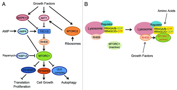
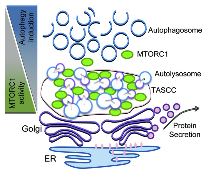
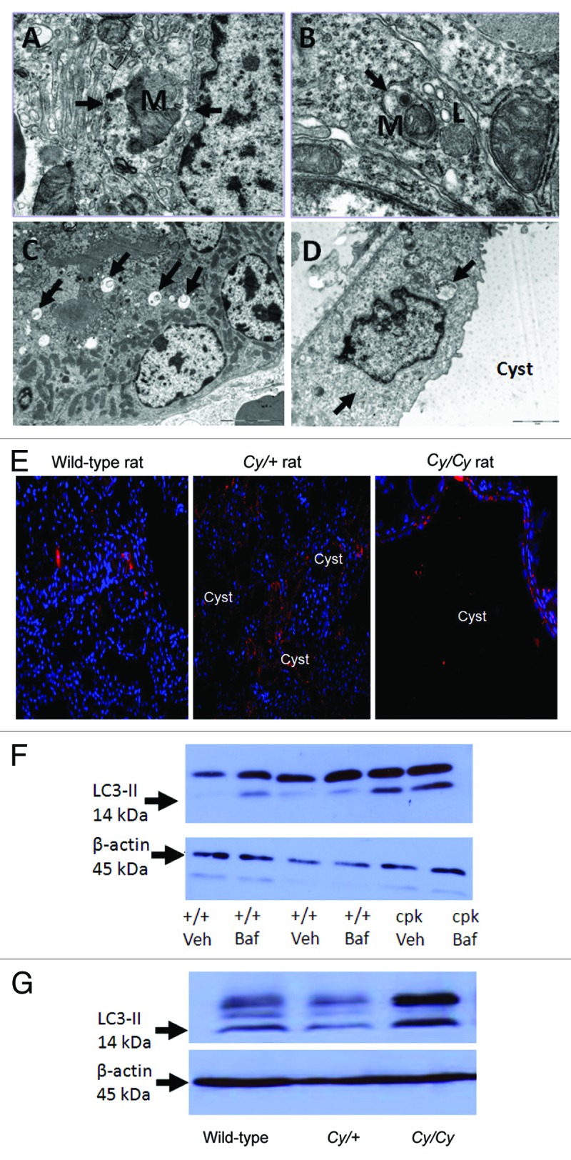
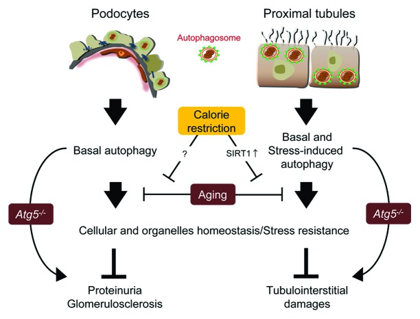
Autophagy is a highly conserved process that degrades cellular long-lived proteins and organelles. Accumulating evidence indicates that autophagy plays a critical role in kidney maintenance, diseases and aging. Ischemic, toxic, immunological, and oxidative insults can cause an induction of autophagy in renal epithelial cells modifying the course of various kidney diseases. This review summarizes recent insights on the role of autophagy in kidney physiology and diseases alluding to possible novel intervention strategies for treating specific kidney disorders by modifying autophagy.

Keywords: acute kidney injury; aging; autophagy; glomerulus; kidney; kidney transplantation; mTOR; podocyte; polycystic kidney disease.


Figures

None
Figure 1. MTOR signaling pathway. (A) Two MTOR complexes (MTORC1 and MTORC2) are stimulated by multiple extra- and intracellular factors. MTORC1 activates translation and cell growth, whereas it inhibits autophagy. (B) Amino acids activate RRAG small GTPases thereby recruiting MTORC1 to the lysosomes where MTORC1 is activated by the RHEB small GTPase.
None
Figure 2. Spatial regulation of MTORC1 and autophagy in podocytes. The proposed functions of the TASCC-like structure in podocytes is depicted. The formation of the TASCC-like structure in podocytes may provide a unique environment producing specific secretory proteins. In the TASCC-like structure, the enriched autolysosomes and lysosomes generate cellular amino acids that may further recruit MTORC1 into the TASCC-like structure via RRAG GTPases. This process creates mutually reciprocal gradients for MTORC1 and autophagic activity within a podocyte, and may allow the cells to convert unnecessary proteins into the special ones that are crucial for keeping or transforming their phenotypes.
None
Figure 3. Autophagy in acute kidney injury. (A) Immunoblot analysis of LC3-II accumulation in kidney tissues after 30 min of bilateral renal ischemia, followed by 0 to 48 h of reperfusion. The figure is adopted from Jiang et al., with permission from Elsevier. (B) Formation of autophagosomes and autolysosomes in tubular cells of kidney tissues during ischemia-reperfusion. Kidney tissues were fixed for EM examination of autophagosomes (left panel, arrows: double or multiple membrane structures containing cytoplasm; middle panel, arrows: undigested organelles such as mitochondria) and autolysosomes (right panel, arrowheads: single membrane structures with remnants of cytoplasmic components). The figure is adopted from Jiang et al., with permission from Elsevier. (C) Cisplatin-induced autophagy in proximal tubular cells in kidney tissues. GFP-LC3 transgenic mice were treated with 20 mg/kg cisplatin for 24 h to harvest kidney tissues for immunostaining of aquaporin-1 (AQP1; marker of proximal tubules). Co-localization of GFP-LC3 dots with AQP1 suggests autophagy induction in proximal tubular cells. The figure is adopted from Inoue et al., with permission from Springer.
None
Figure 4. Autophagy in PKD. Autophagosomes (arrows) are demonstrated in kidneys of +/+ rats (A), Cy/+ rats (B), +/+ mice (C) and in the tubular cells lining the cysts in cpk mice (D). Lysosomes (L) fusing to autophagosomes are shown in (B). Mitochondria (M) within a autophagosome (mitophagy) is shown in (A and B). Reproduced from Belibi et al., with permission from the American Physiological Society. Magnification is X20000 in (A), X40000 in (B), X6000 in (C) and X12000 in (D). (E) To demonstrate that LC3 is present in the cells lining the cysts, IF was performed. There is LC3 staining in cells lining the cysts in Cy/+ and Cy/Cy rats. Nuclei are represented by blue (DAPI) staining. LC3 staining is cytosolic. Reproduced from Belibi et al., with permission from the American Physiological Society. (F) The intensity of the LC3-II bands was increased in cpk mice compared with wild type (+/+) mice. In +/+ mice treated with bafilomycin A1 (+/+ Baf), there was an increase in LC3-II. In cpk mice treated with bafilomycin A1 (cpk Baf), LC3-II was not increased. (G) LC3-II was increased in Cy/Cy rats compared with +/+ and Cy/+ rats. ACTB/β-actin used as a loading control was not different between the groups. Reproduced from Belibi et al., with permission from the American Physiological Society.
None
Figure 5. Autophagy in kidney aging. In both podocytes and proximal tubular cells, autophagy maintains cellular and organelles homeostasis under both basal and stress conditions. Normal aging process or a deletion of Atg5 gene alters autophagy in podocytes and proximal tubular cells, leading to kidney aging. Calorie restriction prevents the progression of kidney aging in a part through SIRT1-dependent autophagy.

References

    1. Mizushima N, Levine B, Cuervo AM, Klionsky DJ. Autophagy fights disease through cellular self-digestion. Nature. 2008;451:1069–75. doi: 10.1038/nature06639. - DOI - PMC - PubMed
    1. Rubinsztein DC. The roles of intracellular protein-degradation pathways in neurodegeneration. Nature. 2006;443:780–6. doi: 10.1038/nature05291. - DOI - PubMed
    1. Yang Z, Klionsky DJ. Eaten alive: a history of macroautophagy. Nat Cell Biol. 2010;12:814–22. doi: 10.1038/ncb0910-814. - DOI - PMC - PubMed
    1. Weide T, Huber TB. Implications of autophagy for glomerular aging and disease. Cell Tissue Res. 2011;343:467–73. doi: 10.1007/s00441-010-1115-0. - DOI - PubMed
    1. Mizushima N, Levine B. Autophagy in mammalian development and differentiation. Nat Cell Biol. 2010;12:823–30. doi: 10.1038/ncb0910-823. - DOI - PMC - PubMed

Publication types