. 2012 Jul;4(7):669-78.
doi: 10.1039/c2mt20025b. Epub 2012 Jun 13.

A structural model of the copper ATPase ATP7B to facilitate analysis of Wilson disease-causing mutations and studies of the transport mechanism

Affiliations

A structural model of the copper ATPase ATP7B to facilitate analysis of Wilson disease-causing mutations and studies of the transport mechanism

Maya Schushan et al. Metallomics. 2012 Jul.

Abstract

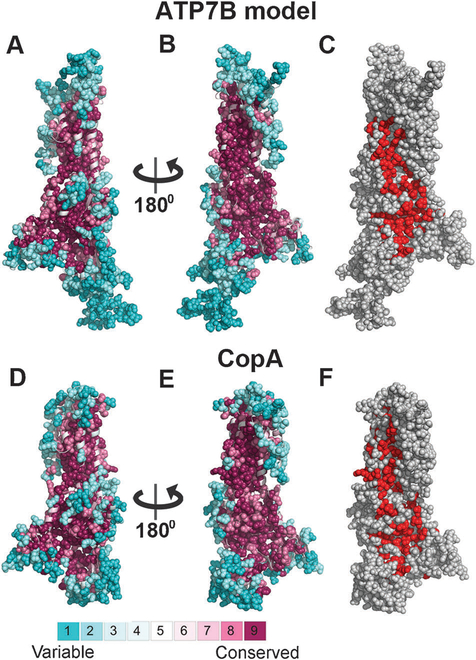
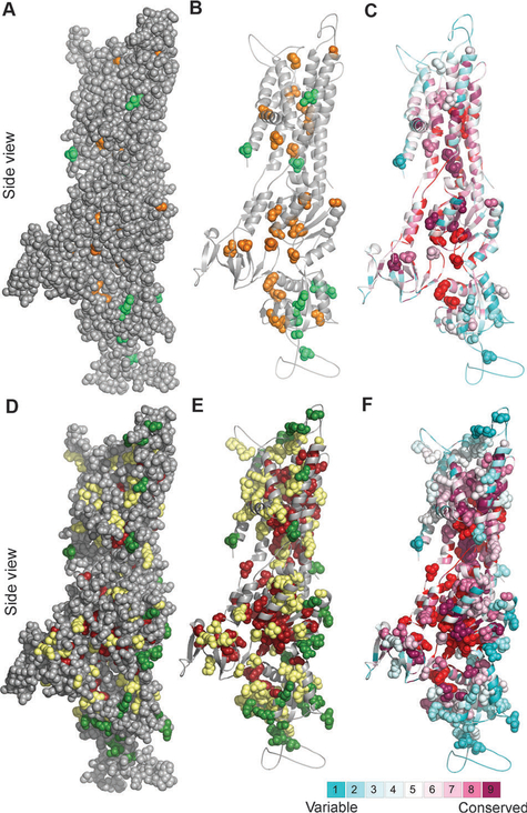
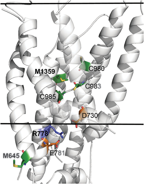
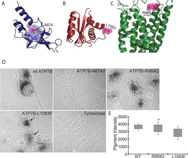
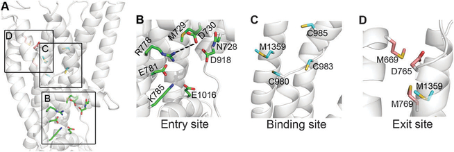
The copper-transporting ATPase ATP7B has an essential role in human physiology, particularly for the liver and brain function. Inactivation of ATP7B is associated with a severe hepato-neurologic disorder, Wilson disease (WD). Hundreds of WD related mutations have been identified in ATP7B to date. The low frequency and the compound-heterozygous nature of causative mutations complicate the analysis of individual mutants and the establishment of genotype-phenotype correlations. To facilitate studies of disease-causing mutations and mechanistic understanding of WD, we have homology-modelled the ATP7B core (residues 643-1377) using the recent structure of the bacterial copper-ATPase LCopA as a template. The model, supported by evolutionary conservation and hydrophobicity analysis, as well as existing and new mutagenesis data, allows molecular interpretations of experimentally characterized clinical mutations. We also illustrate that structure and conservation can be used to grade potential deleterious effects for many WD mutations, which were clinically detected but have not yet been experimentally characterized. Finally, we compare the structural features of ATP7B and LCopA and discuss specific features of the eukaryotic copper pump.


Figures

Fig. 1
Fig. 1. Conservation analysis of ATP7B and LCopA.
Panels A–B and D–E show side-views of the LCopA structure and ATP7B model in cartoon representation, with the cytoplasm below. The structures are colored according to the ConSurf color bar (http://consurf.tau.ac.il), with cyan-to-maroon indicating variable-to-conserved. Positions assigned with the highest or lowest conservation grades (8–9 or 1–2, respectively) are displayed as full atoms spheres, demonstrating the high conservation of the core as opposed to the variability of the peripheral and loop regions. In panels C and F, the LCopA structure and ATP7B model are shown as in panels B and E, respectively, and colored gray, with the functional patches detected by PatchFinder in red. Overall, the conservation patterns of the LCopA and ATP7B are remarkably similar, including the location of a highly conserved patch at the proteins’ surface.
Fig. 2
Fig. 2. Clinically-detected mutations in light of structure and conservation.
Side view of the ATP7B model with the cytoplasm below. (Panels A–C) Location and conservation of clinical missense mutations, for which experimental data are available (Table S1, ESI†) (A) Positions sensitive and non-sensitive to mutation are colored orange and green, respectively. The model (in gray) is shown as spheres, demonstrating which of the experimentally assessed positions are buried in the protein core. (B) Same as in panel A, with only the positions sensitive and non-sensitive to mutation in experiment shown as spheres. (C) As in panel B, with the structure colored by ConSurf analysis (Fig. 1A), and the PatchFinder patch (Fig. 1C) colored red. Most sensitive-to-mutation positions map to conserved residues, either buried or part of the functional site, whereas the less sensitive positions tend to be less conserved and more accessible. (Panels D–F) Location and conservation of all clinical missense mutations (D) Green and dark red coloring indicate positions suggested by our analysis to be sensitive and less sensitive, respectively, positions for which the effect was not proposed are shown in yellow. (E) Same as panel D, showing only the clinical missense mutations as spheres. (F) Same as panel E, with the coloring scheme corresponding to the ConSurf and PatchFinder analysis.
Fig. 3
Fig. 3. Structural location and molecular interactions of M645, R778 and M1359.
The model-structure of ATP7B is shown as white cartoon, and viewed from the side with the cytoplasm below. The view is focused on the membrane domain, and the estimated membrane boundaries, calculated by the PPM webserver (https://opm.phar.umich.edu/ppm_server.php), are marked by the two solid lines. M645, R778, M1359 and interacting residues of interest are shown as sticks, with Met and Cys in green, Arg in blue and Asp and Glu in orange. Nitrogen, Oxygen and Sulfur atoms of these residues are colored blue, red and yellow, respectively. For clarity, the C-termini of TM2 and TM3 are partly transparent. The variable position M645 is situated at the beginning of TM1, facing outwards in contrast to highly conserved M1359 that is situated at the protein core, facing several other potential highly conserved copper ligands (sticks). The conserved R778 is situated at the region corresponding to the suggested copper entry site of LCopA, in close proximity to highly conserved acidic residues, potentially forming stabilizing interactions.
Fig. 4
Fig. 4. Structural location and copper transport activity of the ATP7B mutants.
(A) The A-domain model shown as blue cartoons. A874 is shown as pink atom-spheres, and surrounding residues in blue spheres. (B) The P-domain model in red, with L1083 shown as pink spheres. (C) The membrane domain in green, with R969 as pink spheres. (D) Development of black pigment by tyrosinase is indicative of copper transport by ATP7B. ATP7B-A874V shows no copper transport activity. ATP7B-R969Q shows activity/intensity of color similar to wtATP7B. ATP7B-L1083F shows variable color intensity as indicated by arrows. Cells transfected with tyrosinase plasmids will not develop color and serve as a negative control for the assay. (E). Quantitation of average pigment intensity using ImageJ.
Fig. 5
Fig. 5. Potential functional sites in the membrane region.
The ATP7B model is shown as transparent white cartoons, and viewed from the side with the inward side facing below. (A) Focus on the membrane domain, with residues of the functional site (Table 1) shown as colored sticks. The three sites, i.e. entry, intra-membrane binding and exit sites, are marked by squares. (B) The entry site, with suggested functional residues as green sticks. R778 and D730 could be salt-bridged. (C) The binding site, with TM3 removed for clarity. The suggested binding site residues (colored cyan) are C983, C985 and Met1359 with C980 playing an assisting role. (D) Exit site residues in pink, along with Met1359. TM2 and TM3 were extracted for clarity.

References

    1. Lutsenko S, Barnes NL, Bartee MY and Dmitriev OY, Function and regulation of human copper-transporting ATPases, Physiol. Rev, 2007, 87(3), 1011–1046. - PubMed
    1. El Meskini R, Culotta VC, Mains RE and Eipper BA, Supplying copper to the cuproenzyme peptidylglycine alpha-amidating mono-oxygenase, J. Biol. Chem, 2003, 278(14), 12278–12284. - PubMed
    1. Setty SR, et al., Cell-specific ATP7A transport sustains copper-dependent tyrosinase activity in melanosomes, Nature, 2008, 454, 1142–1146. - PMC - PubMed
    1. Ashino T, et al., Unexpected role of the copper transporter ATP7A in PDGF-induced vascular smooth muscle cell migration, Circ. Res, 2010, 107(6), 787–799. - PMC - PubMed
    1. Das SK and Ray K, Wilson’s disease: an update, Nat. Clin. Pract. Neurol, 2006, 2(9), 482–493. - PubMed

Publication types

MeSH terms

LinkOut - more resources