. 2012 Aug 17;287(34):28852-64.
doi: 10.1074/jbc.M112.364182. Epub 2012 Jun 12.

Metabolism of vertebrate amino sugars with N-glycolyl groups: mechanisms underlying gastrointestinal incorporation of the non-human sialic acid xeno-autoantigen N-glycolylneuraminic acid

Affiliations

Metabolism of vertebrate amino sugars with N-glycolyl groups: mechanisms underlying gastrointestinal incorporation of the non-human sialic acid xeno-autoantigen N-glycolylneuraminic acid

Kalyan Banda et al. J Biol Chem. .

Abstract

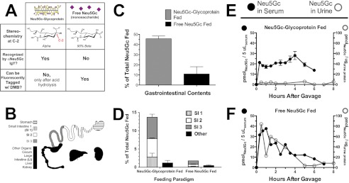
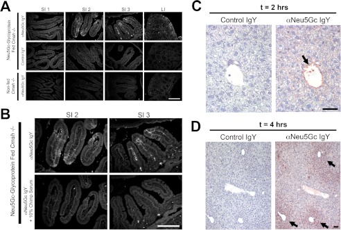
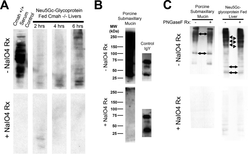
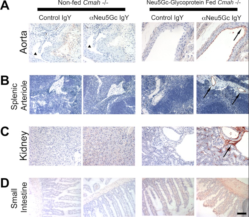
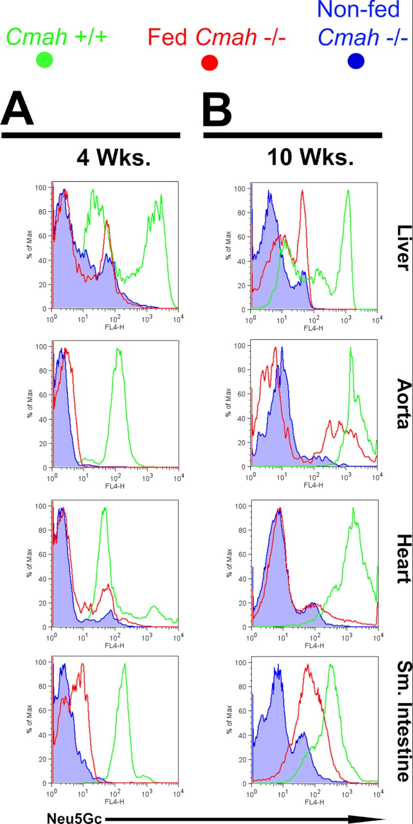
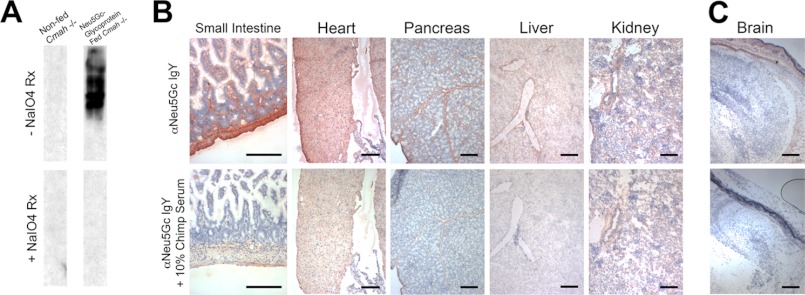
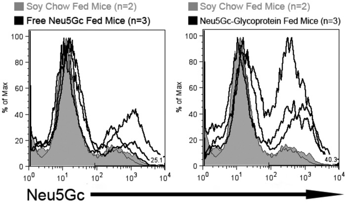
Although N-acetyl groups are common in nature, N-glycolyl groups are rare. Mammals express two major sialic acids, N-acetylneuraminic acid and N-glycolylneuraminic acid (Neu5Gc). Although humans cannot produce Neu5Gc, it is detected in the epithelial lining of hollow organs, endothelial lining of the vasculature, fetal tissues, and carcinomas. This unexpected expression is hypothesized to result via metabolic incorporation of Neu5Gc from mammalian foods. This accumulation has relevance for diseases associated with such nutrients, via interaction with Neu5Gc-specific antibodies. Little is known about how ingested sialic acids in general and Neu5Gc in particular are metabolized in the gastrointestinal tract. We studied the gastrointestinal and systemic fate of Neu5Gc-containing glycoproteins (Neu5Gc-glycoproteins) or free Neu5Gc in the Neu5Gc-free Cmah(-/-) mouse model. Ingested free Neu5Gc showed rapid absorption into the circulation and urinary excretion. In contrast, ingestion of Neu5Gc-glycoproteins led to Neu5Gc incorporation into the small intestinal wall, appearance in circulation at a steady-state level for several hours, and metabolic incorporation into multiple peripheral tissue glycoproteins and glycolipids, thus conclusively proving that Neu5Gc can be metabolically incorporated from food. Feeding Neu5Gc-glycoproteins but not free Neu5Gc mimics the human condition, causing tissue incorporation into human-like sites in Cmah(-/-) fetal and adult tissues, as well as developing tumors. Thus, glycoproteins containing glycosidically linked Neu5Gc are the likely dietary source for human tissue accumulation, and not the free monosaccharide. This human-like model can be used to elucidate specific mechanisms of Neu5Gc delivery from the gut to tissues, as well as general mechanisms of metabolism of ingested sialic acids.


Figures

FIGURE 1.
FIGURE 1.
Different gastrointestinal handling of Neu5Gc from free Neu5Gc or Neu5Gc-glycoprotein feeding. A, figure shows the two feeding strategies compared (Neu5Gc-glycoprotein, left column; free Neu5Gc, right column) and the two key reagents used in this study. αNeu5Gc IgY can recognize glycosidically bound Neu5Gc, such as on PSM (“Neu5Gc-glycoprotein” in this study). DMB is a fluorogenic tag that can react only with free Neu5Gc monosaccharide for quantification in HPLC. Acid hydrolysis can be used to release glycosidically linked Neu5Gc from glycans so that it can be tagged by DMB and is an important process step for quantifying total Neu5Gc (with acid hydrolysis) and free Neu5Gc (without acid hydrolysis). B, schematic of tissues studied in this paper. The contents from stomach and small and large intestines were collected. The small intestine was divided into three isometric sections (SI1, SI2, and SI3), along with the samples collected from stomach, large intestine (LI), liver, and kidney. Urine and feces were also collected. C, Neu5Gc recovered from contents of the gastrointestinal tract (contents from stomach and small and large intestines) was expressed as a percentage of amount gavaged (0.3 mmol of Neu5Gc). We recovered significantly more Neu5Gc from the content of Neu5Gc-glycoprotein-fed mice than free Neu5Gc-fed mice. D, marked differences between Neu5Gc recovered from organs of free Neu5Gc and Neu5Gc-glycoprotein-fed mice at 2 h after feeding. SI1 (hatched bar), SI2 (white bar), SI3 (gray bar), and others (black bar): liver, kidney, and cecum/LI. Neu5Gc-glycoprotein feeding leads to Neu5Gc being retained in the intestines longer, compared with rapid absorption of free Neu5Gc from the intestinal contents. Neu5Gc from Neu5Gc-glycoprotein feeding is particularly enriched in the terminal small intestine (SI3, gray bar). E, top panel, blood and urinary excretory kinetics of Neu5Gc in Neu5Gc-glycoprotein-fed mice. Neu5Gc maintains a near steady state for up to 6 h after feeding in blood (solid black circles) and is minimally excreted in urine (open gray circles) even at the end of 48 h (data not shown). Neu5Gc was very minimally detected in feces from Neu5Gc-glycoprotein-fed mice, ruling it out as major means of excretion. Neu5Gc derived from glycoprotein sources is unlikely to be excreted unchanged in large amounts. Bottom panel, free Neu5Gc-fed mice show a spike and rapid disappearance of Neu5Gc from blood (solid black circles), indicating that it is rapidly absorbed from the gastrointestinal tract. Neu5Gc in urine from these mice shows similar kinetics, a spike and rapid disappearance (open gray circles). The scale of the y axes in these panels is the same. Very minimal amounts of free Neu5Gc were detected in feces of free Neu5Gc-fed mice.
FIGURE 2.
FIGURE 2.
Intestinal uptake of Neu5Gc seen only in Neu5Gc-glycoprotein-fed mice. A, frozen sections of indicated intestine from Neu5Gc-glycoprotein-fed mice 2 h after feeding. Sections are stained with 1:5000 αNeu5Gc IgY (top row) showing prominent uptake (white arrows) in the middle (SI2) and terminal (SI3) part of the small intestine, agreeing with Fig. 1D. Control IgY shows absence of staining on same tissues (middle row). Importantly, tissues from non-fed Cmah−/− mice show absence of staining with αNeu5Gc IgY (bottom row). B, to control for the staining in A, we repeated staining in SI2 and SI3 with and without 10% chimpanzee serum during the primary antibody incubation step. Chimpanzee serum is a rich source of Neu5Gc-containing glycoproteins and blocked staining seen by αNeu5Gc IgY in the SI segments (bottom row). C and D, uptake of Neu5Gc in Neu5Gc-glycoprotein-fed mice by the liver. To determine whether Neu5Gc derived from Neu5Gc-glycoprotein feeding is trafficked to peripheral tissues, we harvested livers of fed mice at 2 h (C) and 4 h (D). Immunohistochemistry using αNeu5Gc IgY revealed staining (black arrows) in the portal vein at 2 h (C), periportal hepatocytes at 4 h (D), and increasingly diffuse periportal hepatocytes at 6 h (figure not shown). Scale bar, 100 μm.
FIGURE 3.
FIGURE 3.
Immunoblots with αNeu5Gc IgY confirm that Neu5Gc from Neu5Gc-glycoprotein feeding is incorporated into endogenous liver glycans. A, immunoblot of Neu5Gc-glycoprotein-fed Cmah−/− liver homogenates (25 μg/lane) at the indicated time point with αNeu5Gc IgY with (bottom) and without (top) periodate treatment (±NaIO4 reaction) of PVDF blotting membrane. Neu5Gc is present on many endogenous liver glycoproteins and signal increases over time. To control for staining, we treated control PVDF membranes with mild periodate (2 mm), which makes Neu5Gc unrecognizable to αNeu5Gc IgY. The positive control, Cmah+/+ Serum Control (left lane), is strong, but 100% sensitive to mild periodate treatment (bottom blot). B, immunoblot of native porcine submaxillary mucin (“Neu5Gc-glycoprotein,” 100 ng/lane) with αNeu5Gc IgY, with (bottom) and without (top) mild periodate treatment (as in A). PSM runs as a high molecular weight smear, with no apparent distinct bands. Chicken IgY (100 ng) was included to show that mild periodate treatment does not interfere with epitope recognition of proteins in SDS-PAGE. C, immunoblot of Neu5Gc-glycoprotein-fed Cmah−/− liver at 6 h with αNeu5Gc IgY with (bottom) and without (top) periodate treatment. To show that Neu5Gc is bioavailable for endogenous glycan synthesis, we used PNGase-F treatment of liver homogenates. PNGase-F treatment did not remove bands but instead leads to a shift toward a lower apparent molecular weight for several bands (black arrowhead) in +PNGase-F lanes, compared with −PNGase-F lanes. Migration of other bands within the same homogenate was not affected by PNGase-F treatment (two-sided black arrows). PSM controls exhibited no apparent shift with PNGase-F treatment, commensurate with mucin-type O-GalNAc glycans on PSM (B). Rx means reaction.
FIGURE 4.
FIGURE 4.
Long term Neu5Gc-glycoprotein feeding leads to metabolic incorporation of Neu5Gc with a human-like tissue distribution I. A–D, paraffin-embedded sections of aorta (A), spleen (B), kidney (C), and small intestine (D) of 3-week Neu5Gc-glycoprotein-fed mice were stained with 1:5000 control IgY or αNeu5Gc IgY (two right columns). Brown staining (see black arrows) with αNeu5Gc IgY, which is not seen with Control IgY, indicates that Neu5Gc is incorporated into these tissues. Similar sections from non-fed Cmah−/− tissues were also stained with control IgY or αNeu5Gc IgY (two left columns), which showed no staining. Collapsed lumen of non-fed aorta (A, left columns) is marked (see black arrowhead). Scale bar, 100 μm.
FIGURE 5.
FIGURE 5.
Long term Neu5Gc-glycoprotein feeding leads to metabolic incorporation of Neu5Gc with a human-like tissue distribution II. A and B, Cmah−/− mice were fed with Neu5Gc-glycoprotein for 3 weeks (A) or 10 weeks (B). At sacrifice, the animals were perfused with collagenase, and organs were dissociated and stained with αNeu5Gc IgY. Flow cytometric analysis showed positive staining for Neu5Gc in Neu5Gc-glycoprotein-fed mice (red traces) above levels seen with non-fed Cmah−/− controls (blue traces) at 4 weeks of feeding (A) and increased further at 10 weeks of feeding (B). Most Neu5Gc-positive events from heart, aorta, and small intestine co-stained with a marker for endothelium (CD31), although Neu5Gc-positive events in the liver co-stained with a marker for hepatocytes (albumin). Tissues from Cmah+/+mice were used as positive controls and for comparison (green traces). These results are representative results from Neu5Gc feeding courses that were repeated at least three times on Cmah−/− animals, staining three Neu5Gc-glycoprotein-fed mice per feeding time point.
FIGURE 6.
FIGURE 6.
Maternal dietary Neu5Gc is metabolically incorporated in utero in newborn tissues. A, Western blot analysis with αNeu5Gc IgY demonstrates Neu5Gc on multiple glycoproteins of fetal small intestinal homogenates (top) that is sensitive to periodate treatment (+NaIO4 reaction, bottom). B, immunohistochemistry with αNeu5Gc IgY of multiple tissues from in utero loaded Cmah−/− newborns indicated widespread incorporation of dietary Neu5Gc, in patterns that exceed those seen with adult feeding. In utero loaded mice demonstrate staining with αNeu5Gc IgY many tissues, including heart, smooth muscle (surrounding intestinal villi), pancreas, liver, and kidney (top row). Staining was controlled with 10% chimpanzee serum block (bottom row). C, consistent with the human condition, we were still unable to load Neu5Gc into the brain, although the cranial and dermis tissue surrounding the brain show staining with αNeu5Gc IgY (top row) that is sensitive to block with chimpanzee serum (bottom row). Scale bar, 100 μm.
FIGURE 7.
FIGURE 7.
Dietary Neu5Gc is incorporated in growing tumors in vivo. To determine whether dietary Neu5Gc can be incorporated in tumors in vivo, adult Cmah−/− mice were injected subcutaneously with 0.5·106 MC38 cells and fed no Neu5Gc (soy chow fed, tinted gray histograms, n = 2), fed free Neu5Gc (solid line in the left histogram, n = 3), or fed glycosidically bound Neu5Gc (solid line in right histogram, n = 3) for 3 weeks. Tumors were removed and digested into single cell suspensions using collagenase. 106 tumor cells from each mouse were stained αNeu5Gc IgY for flow cytometric analysis. Neu5Gc was incorporated into tumor cells from Neu5Gc-glycoprotein-fed mice and free Neu5Gc-fed mice, although at lower levels as indicated by the lower intensity of staining.

References

    1. Schauer R. (1978) Characterization of sialic acids. Methods Enzymol. 50, 64–89 - PubMed
    1. Troy F. A. (1992) Polysialylation. From bacteria to brains. Glycobiology 2, 5–23 - PubMed
    1. Varki A., Schauer R. (2009) in Essentials of Glycobiology (Varki A., Cummings R. D., Esko J. D., Freeze H. H., Stanley P., Bertozzi C. R., Hart G. W., Etzler M. E., eds) pp. 199–218, Cold Spring Harbor Laboratory Press, Cold Spring Harbor, NY - PubMed
    1. Schwarzkopf M., Knobeloch K. P., Rohde E., Hinderlich S., Wiechens N., Lucka L., Horak I., Reutter W., Horstkorte R. (2002) Sialylation is essential for early development in mice. Proc. Natl. Acad. Sci. U.S.A. 99, 5267–5270 - PMC - PubMed
    1. Shaw L., Schauer R. (1988) The biosynthesis of N-glycoloylneuraminic acid occurs by hydroxylation of the CMP-glycoside of N-acetylneuraminic acid. Biol. Chem. Hoppe-Seyler 369, 477–486 - PubMed

Publication types

LinkOut - more resources