. 2012 Nov;92(5):939-50.
doi: 10.1189/jlb.0312122. Epub 2012 Jun 12.

IFN-γ-independent intraocular tumor rejection is mediated by a macrophage-dependent process that leaves the eye intact

Affiliations

IFN-γ-independent intraocular tumor rejection is mediated by a macrophage-dependent process that leaves the eye intact

Terry G Coursey et al. J Leukoc Biol. 2012 Nov.

Abstract

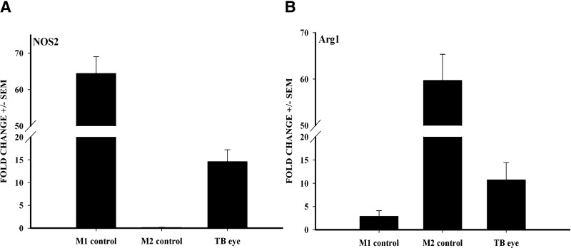
Intraocular tumors reside in an immune-privileged site, yet in certain circumstances, they can undergo immune rejection. Ocular tumor rejection can follow one of two pathways. One pathway is CD4(+) T cell-dependent and culminates in ischemic necrosis of the tumor and phthisis (atrophy) of the eye. A second pathway is also CD4(+) T cell-dependent but does not inflict collateral injury to ocular tissues, and the eye is preserved. We isolated two clones of a murine tumor, Ad5E1 that undergo profoundly different forms of immune rejection in the eye. Clone 2.1 tumors undergo an ischemic necrotizing form of rejection that requires IFN-γ, T cells, and ocular macrophages and culminates in destruction of the eye. By contrast, the second clone of Ad5E1, clone 4, undergoes rejection that also requires T cells and ocular macrophages, but leaves the eye in pristine condition (nonphthisical rejection). Here, we demonstrate that nonphthisical tumor rejection of clone 4 tumors is IFN-γ-independent but requires an ocular macrophage population that contains M1 and M2 macrophages. Clone 4 tumor-bearing eyes displayed ten- and 15-fold increases in M2- and M1-associated markers Arg1 and NO2, respectively. This is in sharp contrast to previous results with clone 2.1 tumor rejection, in which M2 markers were undetectable, and the eye was destroyed. These results suggest that the presence of M2 macrophages tempers the immune rejection of intraocular tumors and promotes immune effectors that inflict minimal injury to innocent bystander cells and thereby preserve the integrity and function of the eye.


Figures

Figure 1.
Figure 1.. Nonphthisical rejection of Ad5E1 clone 4 ocular tumors is immune-mediated and requires T cells.
(A) Normal architecture of a mouse eye after nonphthisical rejection of Ad5E1 clone 4 ocular tumors on Day 21. (B) Normal C57BL/6 mouse eye. (C) Phthisical rejection of Ad5E1 clone 2.1 ocular tumors at Day 21. (D) Progressive growth of intraocular Ad5E1 clone 4 tumors in SCID C57BL/6 mice and rejection in WT C57BL/6 mice. Ad5E1 clone 4 tumor cells (3×10⁵ cells/6 μl) were injected into the AC on Day 0. Tumor growth was scored as the percentage of AC occupied by tumor. C, Cornea; L, lens. Graph represents the combined results of three independent experiments (n=5 mice/group/experiment=a total of 15 mice for each group in the three experiments).
Figure 2.
Figure 2.. Nonphthisical rejection of Ad5E1 clone 4 tumors is mediated by CD8+ or CD4+ T cells.
(A) C57BL/6 mice were depleted of CD8+ T cells or were untreated prior to AC injection of 3 × 10⁵ Ad5E1 clone 4 tumor cells. (B) C57BL/6 CD4 KO or WT mice were injected with 3 × 10⁵ Ad5E1 clone 4 tumor cells. Tumor growth was scored as the percentage of AC occupied by tumor. Graph represents the combined results of three independent experiments (n=5 mice/group/experiment=a total of 15 mice for each group in the three experiments).
Figure 3.
Figure 3.. Nonphthisical rejection of Ad5E1 clone 4 tumors does not require IFN-γ.
(A) C57BL/6 IFN-γ KO or WT mice were AC-injected with 3 × 10⁵ Ad5E1 clone 4 tumor cells. Tumor growth was scored as the percentage of AC occupied by tumor. Graph represents the combined results of three independent experiments (n=5 mice/group/experiment=a total of 15 mice for each group in the three experiments). (B) Ad5E1 clone 4 tumor cells were stained with anti-IFN-γR (open histogram) or an isotype control antibody (shaded histogram) and were evaluated by flow cytometry.
Figure 4.
Figure 4.. Macrophages are necessary for rejection of intraocular Ad5E1 clone 4 tumors and mediate tumor cytotoxicity.
(A) C57BL/6 mice were injected in the AC with 3 × 10⁵ Ad5E1 clone 4 tumor cells. Following tumor injection, mice were injected SCJ with C12MDP-LIP or PBS-LIP every 3–4 days. Ad5E1 tumors were rejected in naïve and PBS-LIP-treated mice but grew progressively in C12MDP-LIP-treated mice. Graph represents the combined results of two independent experiments (n=10 mice/group/experiment=a total of 20 mice/group for the two experiments). (B) BMDMs were untreated or treated with IFN-γ and activated with LPS. BMDMs were then cocultured with 3H-thymidine-labeled Ad5E1 clone 4 tumor cells or B16F10 melanoma cells (unrelated tumor target) at a 10:1 E:T ratio for 48 h, and the percent tumor cell cytotoxicity was determined. Results are expressed as mean ± sem. These experiments were performed three times with similar results.
Figure 5.
Figure 5.. Macrophage-mediated cytotoxicity of Ad5E1 clone 4 tumor cells is partially contact-dependent.
3H-Thymidine-labeled Ad5E1 clone 4 tumors cells (1×10⁴) were plated in the bottom chamber of a transwell culture plate. Resting or activated BMDMs (1×10⁵) were seeded in the upper chamber. Cells were cultured for 48 h, and the percent of tumor cell cytotoxicity was determined. Results are expressed as mean ± sem. This experiment was performed twice with similar results.
Figure 6.
Figure 6.. Ad5E1 clone 4 intraocular tumors have M1 and M2 tumor-infiltrating macrophages.
(A) C57BL/6 mice bearing Ad5E1 clone 4 tumors [tumor-bearing (TB)] were sacrificed 14 days post-tumor injection, and tumor-bearing eyes and control naïve eyes were harvested and homogenized. RAW 264.7 macrophage cells were polarized into an M1 or M2 phenotype by culturing with IFN-γ/LPS or IL- 4/IL-10/IL-13 and were used as positive controls for M1 and M2 cells, respectively. RNA was isolated, and qPCR was performed to determine the expression of NOS2 (A) or Arg1 (B). Samples were compared with a naïve eye and normalized to GAPDH. Results are expressed as mean ± sem. This experiment was performed twice with similar results (n = 5 mice/group for a total of 10 mice/group for the combined experiments).
Figure 7.
Figure 7.. Macrophages mediate killing of Ad5E1 clone 4 tumor cells by soluble factors between 50 kDa and 100 kDa.
(A) Supernatants, from activated macrophages cultured with Ad5E1 clone 4 tumor cells (48 h), were added to 3H-thymidine-labeled Ad5E1 clone 4 tumor cells. Supernatants were fractionated by molecular weight and added to tumor cells. Heat-inactivated AM + T supernatant, activated macrophages plus tumor cell coculture supernatants incubated for 10 min at 100°C, PK=proteinase K-treated AM + T supernatants; PI, proteinase inhibitor-treated AM + T supernatants; ND, none detected. (B) Ad5E1 clone 4 tumors cells were untreated, treated with staurosporine, or treated with AM + T for 48 h. Cells were then stained with annexin V and PI (propidium iodide). Graph represents the percentage of cells that were annexin V-positive. Results are expressed as mean ± sem. This experiment was performed twice with similar results.
Figure 8.
Figure 8.. Macrophages mediate killing of Ad5E1 clone 4 tumor cells by proteases.
Supernatants, from activated macrophages cultured with Ad5E1 clone 4 tumor cells for 48 h, were added to 3H-thymidine-labeled Ad5E1 clone 4 tumor cells. Protease inhibitors (pefabloc, leupeptin, pepstatin) were added to respective experimental groups. As a control, labeled Ad5E1 clone 4 tumor cells were treated with a multicategory broad protease inhibitor cocktail. Results are expressed as mean ± sem; *P < 0.05. This experiment was performed twice with similar results.

Comment in

Similar articles

Cited by

References

    1. Whitsett C. F., Stulting R. D. (1984) The distribution of HLA antigens on human corneal tissue. Invest. Ophthalmol. Vis. Sci. 25, 519–524 - PubMed
    1. Streilein J. W. (2003) Ocular immune privilege: therapeutic opportunities from an experiment of nature. Nat. Rev. Immunol. 3, 879–889 - PubMed
    1. Niederkorn J. Y. (2006) See no evil, hear no evil, do no evil: the lessons of immune privilege. Nat. Immunol. 7, 354–359 - PubMed
    1. Niederkorn J. Y. (2002) Immune privilege in the anterior chamber of the eye. Crit. Rev. Immunol. 22, 13–46 - PubMed
    1. Medawar P. B. (1948) Immunity to homologous grafted skin. III. The fate of skin homografts transplanted to the brain, to subcutaneous tissue, and to the anterior chamber of the eye. Br. J. Exp. Pathol. 29, 58–69 - PMC - PubMed

Publication types

Substances