. 2012 Jun;24(6):2624-34.
doi: 10.1105/tpc.112.099325. Epub 2012 Jun 12.

Demethylesterification of the primary wall by PECTIN METHYLESTERASE35 provides mechanical support to the Arabidopsis stem

Affiliations

Demethylesterification of the primary wall by PECTIN METHYLESTERASE35 provides mechanical support to the Arabidopsis stem

Shoko Hongo et al. Plant Cell. 2012 Jun.

Abstract

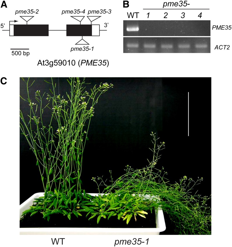
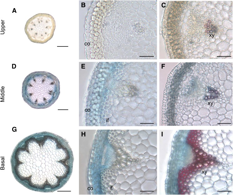
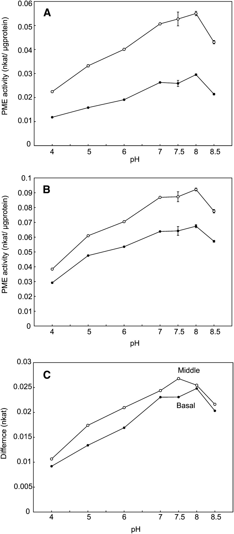
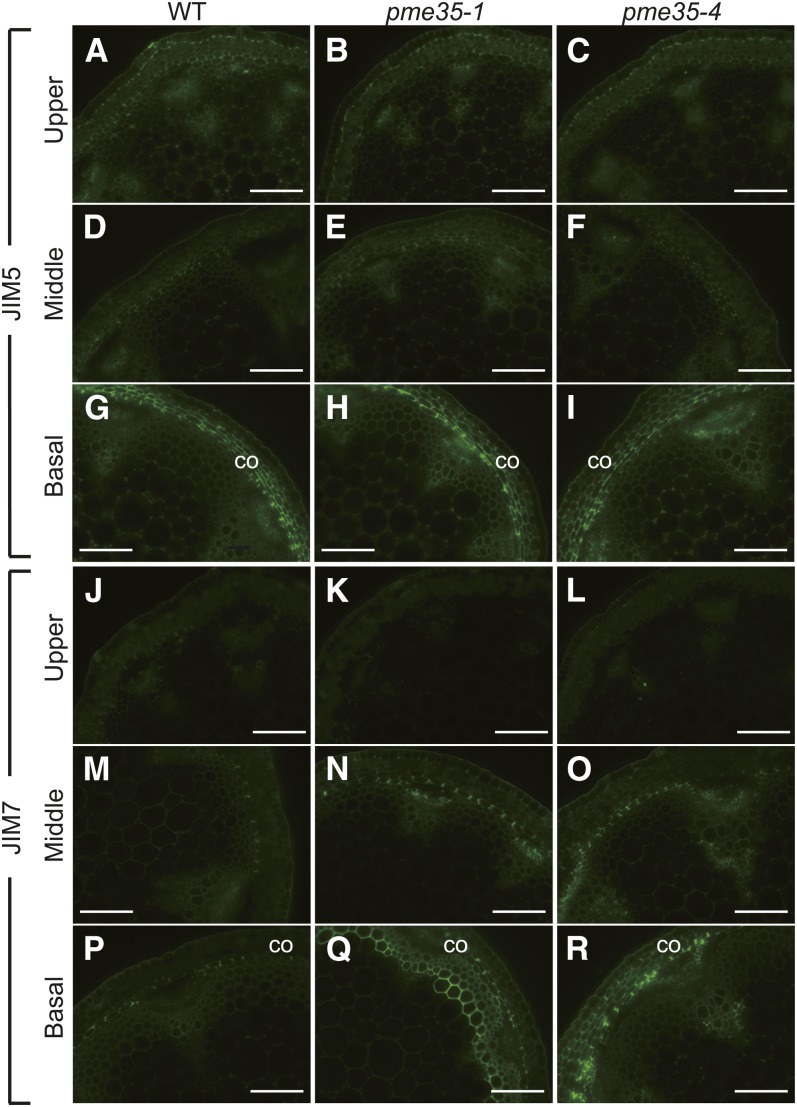
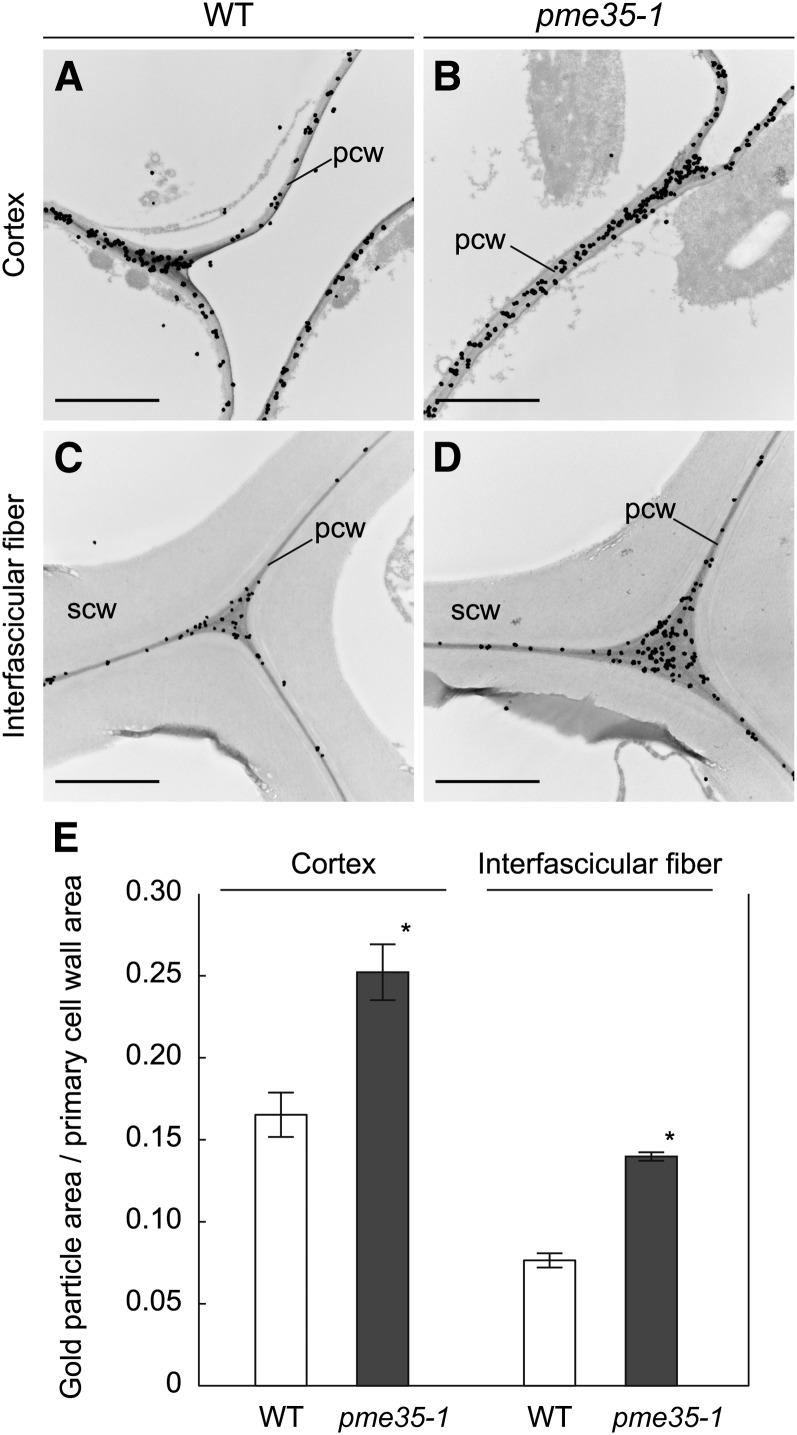
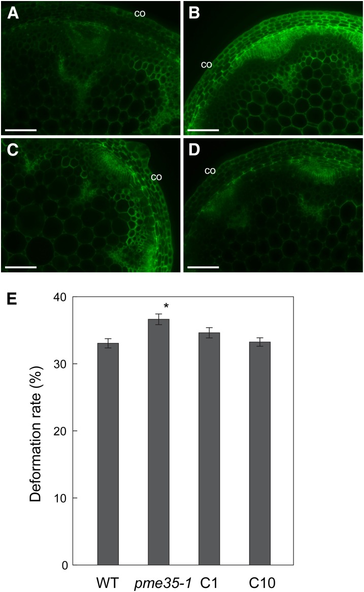
Secondary cell walls, which contain lignin, have traditionally been considered essential for the mechanical strength of the shoot of land plants, whereas pectin, which is a characteristic component of the primary wall, is not considered to be involved in the mechanical support of the plant. Contradicting this conventional knowledge, loss-of-function mutant alleles of Arabidopsis thaliana PECTIN METHYLESTERASE35 (PME35), which encodes a pectin methylesterase, showed a pendant stem phenotype and an increased deformation rate of the stem, indicating that the mechanical strength of the stem was impaired by the mutation. PME35 was expressed specifically in the basal part of the inflorescence stem. Biochemical characterization showed that the activity of pectin methylesterase was significantly reduced in the basal part of the mutant stem. Immunofluorescence microscopy and immunogold electron microscopy analyses using JIM5, JIM7, and LM20 monoclonal antibodies revealed that demethylesterification of methylesterified homogalacturonans in the primary cell wall of the cortex and interfascicular fibers was suppressed in the mutant, but lignified cell walls in the interfascicular and xylary fibers were not affected. These phenotypic analyses indicate that PME35-mediated demethylesterification of the primary cell wall directly regulates the mechanical strength of the supporting tissue.


Figures

Figure 1.
Figure 1.
Structure of PME35 and T-DNA Insertion Sites of pme35 Mutants. (A) T-DNA insertion sites of four allelic mutants of PME35: pme35-1, SALK_019255; pme35-2, SALK_012478; pme35-3, SALK_084661; and pme35-4, SALK_041794. PME35 consists of two exons (black boxes), one intron (black line), and untranslated sequences (white box). (B) PME35 transcript levels as detected by RT-PCR in 35-d-old plants of the wild type (WT) and the four allelic mutants of PME35. ACT2 was used as a loading control. Three biologically independent experiments were performed, and representative data are shown. (C) Forty-day-old plants of the wild type and pme35-1. pme35-1 showed a pendant stem phenotype. Bar = 10 cm.
Figure 2.
Figure 2.
Histochemical Analysis of PME35 Expression and Lignin Deposition Pattern. (A) and (B) Cross sections derived from the upper part of the inflorescence stem of a ProPME35:GUS plant. (C) A GUS-stained section of the upper part of the inflorescence stem of a ProPME35:GUS plant was further stained with phloroglucinol-HCl to disclose lignin deposition in red. (D) and (E) Cross sections derived from the middle part of the inflorescence stem of a ProPME35:GUS plant. (F) A GUS-stained section of the middle part of the inflorescence stem of a ProPME35:GUS plant was further stained with phloroglucinol-HCl. (G) and (H) Cross sections derived from the basal part of the inflorescence stem of a ProPME35:GUS plant. (I) A GUS-stained section of the basal part of the inflorescence stem of a ProPME35:GUS plant was further stained with phloroglucinol-HCl. co, cortex; if, interfascicular fiber; xy, xylem. Bars = 100 µm in (A), (D), and (G) and 20 µm in (B), (C), (E), (F), (H), and (I).
Figure 3.
Figure 3.
PME Activity Assay at Different pH Levels. (A) and (B) PME activity in the middle (A) and basal (B) parts of the inflorescence stem of the wild type and pme35-1 at pH levels of 4, 5, 6, 7, 7.5, 8, and 8.5. Means with se (n = 3) are shown. Open circle, the wild type; closed circle, pme35-1. (C) Differences in PME activity between the wild type and pme35-1 in the middle and basal parts of the inflorescence stem at pH levels of 4, 5, 6, 7, 7.5, 8, and 8.5. Open circle, middle part of the inflorescence stem; closed circle, basal part of inflorescence stem.
Figure 4.
Figure 4.
PME Activity and PME35 Transcriptional Level in the Upper, Middle, and Basal Parts of the Inflorescence Stem. (A) PME activity in the upper, middle, and basal part of the inflorescence stem of the wild type (WT) and pme35-1. Enzyme activity was assayed at pH 7.5. Means with se (n = 3) are shown. Asterisks indicate that the value of pme35-1 was significantly different from that of the wild type at P < 0.05. (B) PME35 consisted of the pro and mature-PME regions. The pro region includes signal peptide sequences, the PME inhibitor domain, and a processing motif. PMEI domain, PME inhibitor domain (gray box); PME domain, PME catalytic domain (black box). Digits above the rectangle indicate the positions of amino acid residues at the beginning and end of each domain. (C) PME35 transcriptional levels in the upper, middle, and basal parts of the inflorescence stem of the wild type were detected by RT-PCR, which were repeated three times independently. CT2 was used as a loading control. Three biologically independent experiments were performed, and representative data are shown.
Figure 5.
Figure 5.
Reduction of PME Activity in pme35 Mutants. PME activity was measured in the wild type (WT) and the four allelic mutants of PME35 in the basal part of the inflorescence stem. Enzyme activity was assayed at pH 7.5. Means with se (n = 3) are shown. Asterisks indicate that the value of the four allelic mutants was significantly different from that of the wild type at P < 0.05.
Figure 6.
Figure 6.
Immunohistochemical Analysis of the Upper to the Basal Part of the Inflorescence Stem of the Wild Type and Two pme35 Mutants. Cross sections derived from the upper ([A] to [C] and [J] to [L]), middle ([D] to [F] and [M] to [O]), and basal ([G] to [I] and [P] to [R]) parts of the inflorescence stem of wild-type (WT; [A], [D], [G], [J], [M], and [P]), pme35-1 ([B], [E], [H], [K], [N], and [Q]), and pme35-4 ([C], [F], [I], [L], [O], and [R]) plants were probed with the JIM5 ([A] to [I]) or JIM7 ([J] to [R]) antibody. co, cortex. Bars = 100 µm.
Figure 7.
Figure 7.
Immunoelectron microscopy of the Basal Part of the Inflorescence Stem. Cross sections derived from the basal part of the inflorescence stem of the wild type and pme35-1 were probed with the JIM7 antibody, and cortical or interfascicular fiber cells were observed. pcw, primary cell wall; scw, secondary cell wall. Bars = 2 µm. (A) Cortical cells of the wild-type (WT) section. (B) Cortical cells of the pme35-1 section. (C) Interfascicular fiber cells of the wild-type section. (D) Interfascicular fiber cells of the pme35-1 section. (E) The degree of gold particle binding was calculated as the gold particle area per primary cell wall area from electron micrographs of cortical or interfascicular fiber cells. Means with se (the number of independent sections used for the counting was 5, 5, 4, and 3 from left to right) are shown. Asterisks indicate that the value of pme35-1 was significantly different from that of the wild type at P < 0.05.
Figure 8.
Figure 8.
Morphology of the Cortical Cell Files and Mechanical Strength in the Basal Part of the Inflorescence Stem. (A) Strength of the basal part of the inflorescence stem as assayed by a compression test. Deformation rate was calculated as the load-induced compressed distance with respect to the diameter of the stem when the applied force reached 2 n. Means with se (n = 20) are shown. Asterisks indicate that the value is significantly different from that of the wild type at P < 0.05 according to Student’s t test. WT, the wild type. (B) Cross sections of the basal part of the inflorescence stem of the wild type were observed by transmission electron microscopy. (C) Cross sections of the basal part of the inflorescence stem of pme35-1 were observed by transmission electron microscopy. (D) Enlarged image from (B). (E) Enlarged images from (C). Boxes in (B) and (C) indicate the regions enlarged in (D) and (E) respectively. co, cortex; if, interfascicular fiber. Bars = 10 µm in (B) and (C) and 2 µm in (D) and (E).
Figure 9.
Figure 9.
Complementation of pme35-1 Phenotypes. Cross sections derived from the basal part of the inflorescence stem of the wild type, pme35-1, and complementation lines of pme35-1 (C1 or C10) were probed with JIM7 antibody and observed under a fluorescence microscope. co, cortex. Bars = 100 µm. (A) Cross section of the wild type. (B) Cross section of pme35-1. (C) Cross section of C1. (D) Cross section of C10. (E) Mechanical strength at the basal part of the inflorescence stem of the wild type (WT), pme35-1, and complementation lines of pme35-1 (C1 or C10). Means with se (n = 20) are shown. Asterisk indicates that the value of pme35-1 or complemented lines was significantly different from that of the wild type at P < 0.05 according to a Student’s t test.

References

    1. Albersheim P., Darvill A., Roberts K., Sederoff R., Staehelin A. (2011). Plant Cell Walls. (New York: Garland Science, Taylor & Francis Group).
    1. Arai-Sanoh Y., et al. (2011). Genotypic variations in non-structural carbohydrate and cell-wall components of the stem in rice, sorghum, and sugar vane. Biosci. Biotechnol. Biochem. 75: 1104–1112 - PubMed
    1. Blumenkrantz N., Asboe-Hansen G. (1973). New method for quantitative determination of uronic acids. Anal. Biochem. 54: 484–489 - PubMed
    1. Bosch M., Hepler P.K. (2005). Pectin methylesterases and pectin dynamics in pollen tubes. Plant Cell 17: 3219–3226 - PMC - PubMed
    1. Bourgault R., Bewley J.D. (2002). Gel diffusion assays for endo-beta-mannanase and pectin methylesterase can underestimate enzyme activity due to proteolytic degradation: A remedy. Anal. Biochem. 300: 87–93 - PubMed

Publication types

MeSH terms

Associated data

LinkOut - more resources