. 2013 Jul;23(7):1703-14.
doi: 10.1093/cercor/bhs165. Epub 2012 Jun 12.

Multivariate activation and connectivity patterns discriminate speech intelligibility in Wernicke's, Broca's, and Geschwind's areas

Affiliations

Multivariate activation and connectivity patterns discriminate speech intelligibility in Wernicke's, Broca's, and Geschwind's areas

Daniel A Abrams et al. Cereb Cortex. 2013 Jul.

Abstract

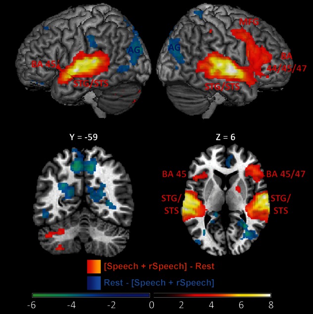
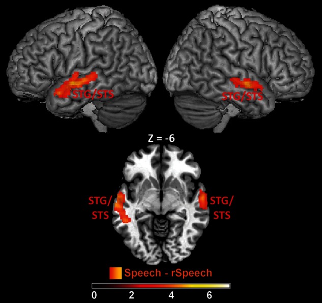
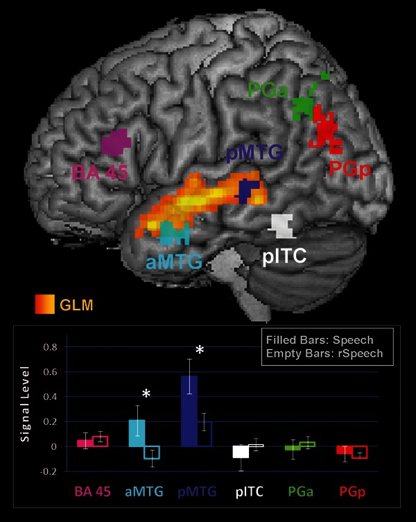
The brain network underlying speech comprehension is usually described as encompassing fronto-temporal-parietal regions while neuroimaging studies of speech intelligibility have focused on a more spatially restricted network dominated by the superior temporal cortex. Here we use functional magnetic resonance imaging with a novel whole-brain multivariate pattern analysis (MVPA) to more fully characterize neural responses and connectivity to intelligible speech. Consistent with previous univariate findings, intelligible speech elicited greater activity in bilateral superior temporal cortex relative to unintelligible speech. However, MVPA identified a more extensive network that discriminated between intelligible and unintelligible speech, including left-hemisphere middle temporal gyrus, angular gyrus, inferior temporal cortex, and inferior frontal gyrus pars triangularis. These fronto-temporal-parietal areas also showed greater functional connectivity during intelligible, compared with unintelligible, speech. Our results suggest that speech intelligibly is encoded by distinct fine-grained spatial representations and within-task connectivity, rather than differential engagement or disengagement of brain regions, and they provide a more complete view of the brain network serving speech comprehension. Our findings bridge a divide between neural models of speech comprehension and the neuroimaging literature on speech intelligibility, and suggest that speech intelligibility relies on differential multivariate response and connectivity patterns in Wernicke's, Broca's, and Geschwind's areas.

Keywords: Angular gyrus; Auditory cortex; Broca's area; Inferior frontal gyrus; Speech perception.


Figures

Figure 1.
Figure 1.
Omnibus responses to Speech and rSpeech. Surface rendering, coronal (Y = − 59), and axial (Z = 6) slices of cortical regions activated during speech and rotated speech conditions (heat map) and deactivated during rest (blue). Images were thresholded using a voxel-wise statistical height threshold of P < 0.005, with corrections for multiple spatial comparisons at the cluster level (P < 0.05). BA 44, pars opercularis of the inferior frontal gyrus; BA 45, pars triangularis of the inferior frontal gyrus; BA 47, pars orbitalis of the inferior frontal gyrus; AG, angular gyrus; STG, superior temporal gyrus; STS, superior temporal sulcus.
Figure 2.
Figure 2.
Univariate responses to Speech versus rSpeech. Surface rendering and axial (Z = − 6) slice of cortical regions with greater activation during intelligible relative to unintelligible speech (the [Speech – rSpeech] contrast). Images were thresholded using a voxel-wise statistical height threshold of P < 0.005, with corrections for multiple spatial comparisons at the cluster level (P < 0.05). No voxels survived this criteria for the [rSpeech – Speech] contrast. STG, superior temporal gyrus; STS, superior temporal sulcus.
Figure 3.
Figure 3.
Multivariate pattern analysis of Speech versus rSpeech. Classification maps show brain regions whose activity patterns discriminated between [Speech – Rest] and [rSpeech – Rest] conditions. Maximum classification accuracy ranged from 80% to 85% across all brain regions identified by MVPA. AG, angular gyrus; FP, frontal pole; aMTG, anterior middle temporal gyrus; pMTG, posterior middle temporal gyrus; BA 45, pars triangularis of the inferior frontal gyrus.
Figure 4.
Figure 4.
Overlap between univariate and multivariate responses to Speech versus rSpeech. Anterior and posterior left middle temporal gyrus (MTG) regions showed significant overlap in multivariate and univariate responses to speech intelligibility. Outside the left MTG, there was no overlap between the MVPA and univariate results. aMTG, anterior middle temporal gyrus; pMTG, posterior middle temporal gyrus; AG, angular gyrus.
Figure 5.
Figure 5.
Signal levels in left-hemisphere ROIs. Signal level differences for Speech and rSpeech conditions are plotted in the bar graph for 6 left-hemisphere regions identified by MVPA. The filled and empty bars in the bar graph represent signal levels for the Speech and rSpeech conditions, respectively. Signal-level differences in the anterior and posterior MTG showed significant signal-level differences between Speech and rSpeech; the pars triangularis of the inferior frontal gyrus (BA 45), the posterior inferior temporal cortex (pITC), and the anterior (PGa) and posterior (PGp) regions of the angular gyrus did not differ in signal level.
Figure 6.
Figure 6.
Functional connectivity during Speech and rSpeech. Top: The locations of spherical 6 mm ROIs used in the functional connectivity analysis are plotted. The center of these spheres is the voxel associated with peak classification accuracy measured with MVPA within each of these brain regions. Middle: Correlation values from the functional connectivity analysis measured in response to the Speech condition. Bottom: Correlation values from the functional connectivity analysis measured in response to the rSpeech condition. Overall functional connectivity was greater in the Speech, compared with the rSpeech conditions. Functional connectivity results that met the criteria for significance (P < 0.005) are labeled with *. BA 45, pars triangularis of the inferior frontal gyrus; PGa, anterior angular gyrus; PGP, posterior angular gyrus; aMTG, anterior middle temporal gyrus; pMTG, posterior middle temporal gyrus.

References

    1. Abrams DA, Bhatara A, Ryali S, Balaban E, Levitin DJ, Menon V. Decoding temporal structure in music and speech relies on shared brain resources but elicits different fine-scale spatial patterns. Cereb Cortex. 2011;21:1507–1518. - PMC - PubMed
    1. Adank P, Devlin JT. On-line plasticity in spoken sentence comprehension: adapting to time-compressed speech. Neuroimage. 2010;49:1124–1132. - PMC - PubMed
    1. Amunts K, Lenzen M, Friederici AD, Schleicher A, Morosan P, Palomero-Gallagher N, Zilles K. Broca's region: novel organizational principles and multiple receptor mapping. PLoS Biol. 2010;8:1–16. - PMC - PubMed
    1. Azadpour M, Balaban E. Phonological representations are unconsciously used when processing complex, non-speech signals. PLoS One. 2008;3:1–7. - PMC - PubMed
    1. Bates E, Wilson SM, Saygin AP, Dick F, Sereno MI, Knight RT, Dronkers NF. Voxel-based lesion-symptom mapping. Nat Neurosci. 2003;6:448–450. - PubMed

Publication types