. 2012;8(5):e1002724.
doi: 10.1371/journal.ppat.1002724. Epub 2012 May 31.

The Wor1-like protein Fgp1 regulates pathogenicity, toxin synthesis and reproduction in the phytopathogenic fungus Fusarium graminearum

Affiliations

The Wor1-like protein Fgp1 regulates pathogenicity, toxin synthesis and reproduction in the phytopathogenic fungus Fusarium graminearum

Wilfried Jonkers et al. PLoS Pathog. 2012.

Abstract

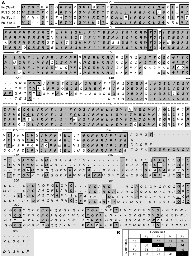
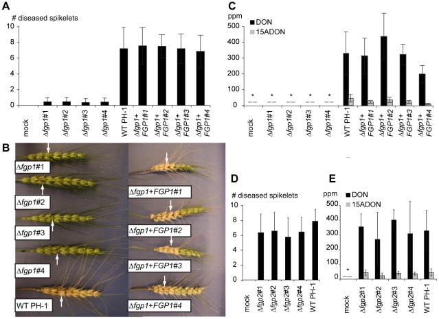
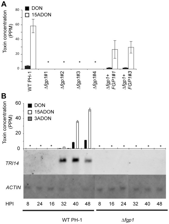
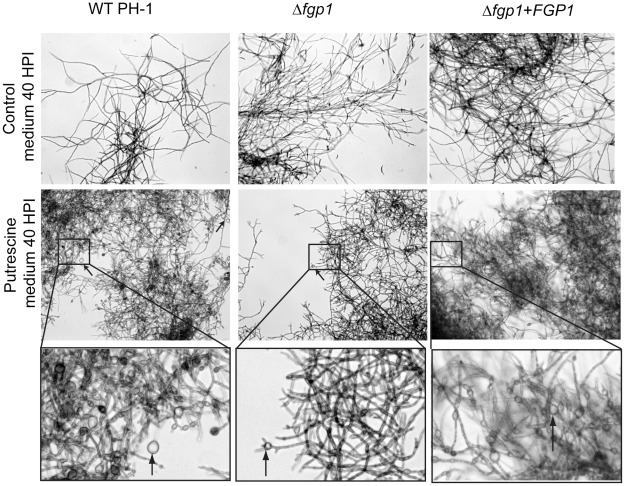
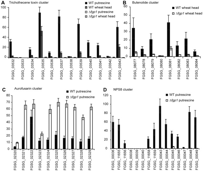
WOR1 is a gene for a conserved fungal regulatory protein controlling the dimorphic switch and pathogenicity determents in Candida albicans and its ortholog in the plant pathogen Fusarium oxysporum, called SGE1, is required for pathogenicity and expression of key plant effector proteins. F. graminearum, an important pathogen of cereals, is not known to employ switching and no effector proteins from F. graminearum have been found to date that are required for infection. In this study, the potential role of the WOR1-like gene in pathogenesis was tested in this toxigenic fungus. Deletion of the WOR1 ortholog (called FGP1) in F. graminearum results in greatly reduced pathogenicity and loss of trichothecene toxin accumulation in infected wheat plants and in vitro. The loss of toxin accumulation alone may be sufficient to explain the loss of pathogenicity to wheat. Under toxin-inducing conditions, expression of genes for trichothecene biosynthesis and many other genes are not detected or detected at lower levels in Δfgp1 strains. FGP1 is also involved in the developmental processes of conidium formation and sexual reproduction and modulates a morphological change that accompanies mycotoxin production in vitro. The Wor1-like proteins in Fusarium species have highly conserved N-terminal regions and remarkably divergent C-termini. Interchanging the N- and C- terminal portions of proteins from F. oxysporum and F. graminearum resulted in partial to complete loss of function. Wor1-like proteins are conserved but have evolved to regulate pathogenicity in a range of fungi, likely by adaptations to the C-terminal portion of the protein.


Conflict of interest statement

The authors have declared that no competing interests exist.

Figures

Figure 1
Figure 1. Alignment of four Wor1-like Fusarium orthologs.
A) Protein sequence alignment of four Fusarium Wor1-like proteins: Fo: Sge1 (FOXG_10510) from F. oxysporum, FVEG_09150 from F. verticillioides, Fg: Fgp1 (FGSG_12164) from F. graminearum and Fs: Fs_81912 from F. solani (Nectria haematococca). Conserved and similar residues are shaded grey. The boxed threonine residue at position 68 is a conserved putative phosphorylation site. The solid black line represents the WOPRa box and the dashed black line the WOPRb box. The C-terminal part is indicated by grey background. The protein alignment was created using MacVector version 10.6.0. B) Table in which the percentages of similarities are given for each of the Wor1-like ortholog (Fg: Fgp1 (FGSG_12164) from F. graminearum, Fv: FVEG_09150 from F. verticillioides, Fo: Sge1 (FOXG_10510) from F. oxysporum and Fs: Fs_81912 from F. solani (Nectria haematococca)) to each of the other protein in the alignment presented in A. Different percentages are presented for the N-terminal portion (white cells) and the C-terminal portion (grey cells).
Figure 2
Figure 2. FGP1 is required for pathogenicity and trichothecene accumulation but FGP2 is not.
A) Average number of diseased spikelets of 30 plants after mock inoculated with H2O, inoculated with four independent FGP1 deletion mutants, with wild type PH-1 or with four independent FGP1 complemented transformants. Wheat heads were point inoculated and disease spread through adjacent spikelets was enumerated after 14 days. Error bars indicate standard deviation. B) Photographs from wild type inoculated wheat head (lower head on the left), wheat heads inoculated with the four FGP1 deletion mutants (upper four heads on the left) and wheat heads inoculated with the four FGP1 complemented transformants (four heads on the right). Arrows indicate the inoculated spikelet in each head photographed. C) DON and 15-ADON concentrations in the inoculated spikelet 14 days after inoculation with H2O, four independent FGP1 deletion mutants, wild type PH-1 or four FGP1 complemented transformants. Asterisks mean no toxin was detected. D) Average number of diseased spikelets of 30 plants after mock inoculated with H2O or inoculated with four independent FGP2 deletion mutants or wild type PH-1. Wheat heads were point inoculated and spread through adjacent spikelets was enumerated after 14 days. Error bars indicate standard deviation. E) DON and 15-ADON concentrations in the inoculated spikelet 14 days after inoculation with H2O, four independent FGP2 deletion mutants or wild type PH-1. Asterisk means no toxin was detected.
Figure 3
Figure 3. The Δfgp1 strain is unable to pass through the rachis node.
The infection behavior of the FGP1 and FGP2 deletion mutants in the wheat head was determined by light and fluorescence microscopy. Spikelets were inoculated and assessed after two to three days for fungal spread within the floral tissue. A) The palea and lemma of a flower inoculated with Δfgp1 show browning. The glume remains green, as no fungal colonization occurs within this portion of the flower. B) The palea, lemma and glume of a flower inoculated with Δfgp2 all show browning. C) The GFP expressing Δfgp1 strain grows inside the flower but does not penetrate the rachis node. Patches of GFP-expressing fungal mycelium are observed along palea and lemma inside the flower (arrow) but no GFP is seen in the rachis node (red circle) or beyond into the rachis. D) The GFP expressing virulent Δfgp2 strain grows in the flower and penetrates the rachis node. Patches of GFP expressing fungal mycelium are observed along palea and lemma inside the flower and GFP is seen within the rachis node (red circle), the rachis and beyond.
Figure 4
Figure 4. Fgp1 is involved in conidiogenesis as well as in ascospore formation.
A) Macroconidia production was assessed in three experiments each in two replicas on mung bean agar (MBA) plates and counted in a haemocytometer. B) Average macroconidium length was determined by measuring 30 macroconidia of each strain. C) Photos of a representative wild type PH-1 macroconidium (left), three representative conidia formed by the FGP1 deletion mutant (middle) and a representative FGP1 complementation strain macroconidium (right). D) Ascospore production by wild type PH-1, two FGP1 deletion mutants and two FGP1 complementation strains was assessed in four replicas for each strain on carrot agar plates two weeks after the mycelium was knocked down using a 2.5% Tween-60 solution. Ascospores were counted with a haemocytometer.
Figure 5
Figure 5. The FGP1 deletion strain does not produce trichothecene toxins in putrescine medium.
A) Histogram of toxin concentration (parts per million - ppm) of DON and 15-ADON measured in putrescine medium after one week of growth of wild type, four Δfgp1 strains and two complemented strains. Asterisks indicate no toxin was detected. B) Histogram of toxin concentration (parts per million - ppm) of DON, 15-ADON and 3-ADON measured in putrescine medium with samples taken at 8, 16, 24, 32, 40, and 48 hours post inoculation (HPI) with wild type PH-1 or a FGP1 deletion strain. Detectable toxin concentrations are found at 32, 40 and 48 HPI in the wild type PH-1 but not in the FGP1 deletion strain at any time point. Asterisks indicate no toxin was detected. A northern blot of RNA obtained from the same samples shows expression of the TRI gene, TRI14, after 32, 40 and 48 HPI in the wild type PH-1 but not in the FGP1 deletion strain at any time point. The loading control gene ACTIN is expressed equally at all time points for both wild type and the FGP1 deletion strain.
Figure 6
Figure 6. Vigorous hyphal swelling during growth in putrescine medium is seen to a lesser extent in Δfgp1.
Mycelium of wild type, a FGP1 deletion strain and a complemented strain growing in control minimal medium (upper panel) or putrescine (middle panel) at 40 HPI. A representative area of the mycelium observed under the microscope is given for each strain. Mycelium of wild type PH-1 and the complemented strain show branching and numerous hyphae with bulbous structures when compared to mycelium grown in control minimal medium. Mycelium of the FGP1 deletion strain also shows branching but fewer hyphae with bulbous structures. Greater magnifications of the boxed frames from the middle pictures (lower panel) show that in the mycelium of the wild type and complemented strains many bulbous structures are formed (left and right pictures in the lower panel). The mycelium of the Δfgp1 strain shows some bulbous structures but less when compared to with wild type and complemented strain (middle picture in the lower panel). Bulbous structures are indicated by arrows in the middle and lower panels.
Figure 7
Figure 7. Fgp1 mediates expression of genes in several gene clusters.
Histograms represent mean relative expression levels from three replicate experiments. Error bars represent standard deviation. A) Expression means for genes from the TRI cluster in wild type PH-1 grown in putrescine (black bars) or during wheat head infection (dark gray bars) and in a FGP1 deletion strain grown in putrescine (white bars) or during wheat head infection (light gray bars). B) Expression means for genes from the butenolide biosynthetic cluster in wild type PH-1 during wheat head infection (black bars) and in a FGP1 deletion strain during wheat head infection (white bars). C) Expression means for genes from the aurofusarin biosynthetic cluster in wild type PH-1 grown in putrescine medium (black bars) or for a FGP1 deletion strain grown in putrescine medium (white bars). D) Expression means for genes from the NPS8 cluster in wild type PH-1 grown in putrescine medium (black bars) or a FGP1 deletion strain grown in putrescine medium (white bars).
Figure 8
Figure 8. FGP1 and SGE1 are not functionally interchangeable between F. graminearum and F. oxysporum.
FGP1 can complement a FGP1 deletion mutant but SGE1 cannot. A chimeric FGP1/SGE1 gene expressed from the FGP1 promoter partially complements a FGP1 deletion mutant. FGP1 cannot complement a SGE1 deletion mutation nor can a chimeric SGE1/FGP1 gene expressed from the SGE1 promoter A) Average number of diseased spikelets of 20–30 wheat plants, two weeks after point inoculation with H2O, a FGP1 deletion mutant, wild type PH-1, one independent FGP1 complementation strain, two independent SGE1 complementation strains and six independent chimeric FGP1/SGE1 strains. B) Average disease index of 20 tomato plants, three weeks after inoculation with H2O, a SGE1 deletion mutant , wild type Fol strain 4287, the original SGE1 complementation strain , two independent FGP1 complementation strains and four independent chimeric SGE1/FGP1 strains.

Similar articles

Cited by

References

    1. Goswami RS, Kistler HC. Heading for disaster: Fusarium graminearum on cereal crops. Mol Plant Pathol. 2004;5:515–525. - PubMed
    1. Trail F. For blighted waves of grain: Fusarium graminearum in the postgenomics era. Plant Physiol. 2009;149:103–110. - PMC - PubMed
    1. Ilgen P, Hadeler B, Maier FJ, Schafer W. Developing kernel and rachis node induce the trichothecene pathway of Fusarium graminearum during wheat head infection. Mol Plant Microbe Interact. 2009;22:899–908. - PubMed
    1. Jansen C, von Wettstein D, Schafer W, Kogel KH, Felk A, et al. Infection patterns in barley and wheat spikes inoculated with wild-type and trichodiene synthase gene disrupted Fusarium graminearum. Proc Natl Acad Sci U S A. 2005;102:16892–16897. - PMC - PubMed
    1. Kimura M, Tokai T, Takahashi-Ando N, Ohsato S, Fujimura M. Molecular and genetic studies of fusarium trichothecene biosynthesis: pathways, genes, and evolution. Biosci Biotechnol Biochem. 2007;71:2105–2123. - PubMed

Publication types

MeSH terms