

doi: 10.1155/2012/403870.
Epub 2012 May 30.
Reactive oxygen species-mediated control of mitochondrial biogenesis
Affiliations
- PMID: 22693510
- PMCID: PMC3369472
- DOI: 10.1155/2012/403870
Item in Clipboard
Reactive oxygen species-mediated control of mitochondrial biogenesis
Int J Cell Biol.
2012.
Abstract
Mitochondrial biogenesis is a complex process. It necessitates the contribution of both the nuclear and the mitochondrial genomes and therefore crosstalk between the nucleus and mitochondria. It is now well established that cellular mitochondrial content can vary according to a number of stimuli and physiological states in eukaryotes. The knowledge of the actors and signals regulating the mitochondrial biogenesis is thus of high importance. The cellular redox state has been considered for a long time as a key element in the regulation of various processes. In this paper, we report the involvement of the oxidative stress in the regulation of some actors of mitochondrial biogenesis.
Figures

The mammalian oxidative phosphorylations (OXPHOS) system. Depicted are the four respiratory complexes (I–IV), electron carriers coenzyme Q and cytochrome c, the ATP synthase complex, the ADP/ATP carrier (ANC); and the phosphate carrier (PiC). Arrows at complexes I, III, and IV illustrate the proton pumping to the intermembrane space. Indicated are the number of complex subunits encoded by mitochondrial (mtDNA) and nuclear (nDNA) genomes.
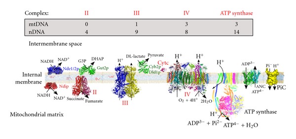
The Saccharomyces cerevisiae oxidative phosphorylations (OXPHOS) system. The main differences with the mammalian OXPHOS system are the absence of complex I that is substituted by external and internal NADH dehydrogenases, and the presence of D, L-lactate dehydrogenases, which transfer electrons directly to cytochrome c. Indicated are the number of protein subunits encoded by mitochondrial (mtDNA) and nuclear (nDNA) genomes.
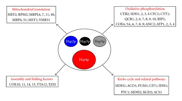
The HAP complex: a master regulator of the mitochondrial biogenesis in the yeast Saccharomyces cerevisiae. The four subunits constituting the complex are represented here. Size differences illustrate the difference in the predicted molecular weights of each subunit. The mitochondrial proteins encoding genes regulated by the complex are also indicated. See text for references.
References
-
- Lowell BB, Shulman GI. Mitochondrial dysfunction and type 2 diabetes. Science. 2005;307(5708):384–387. - PubMed
-
- Terzioglu M, Larsson NG. Mitochondrial dysfunction in mammalian ageing. Novartis Foundation Symposium. 2007;287:197–208. - PubMed
-
- Larsson N-G. Somatic mitochondrial DNA mutations in mammalian aging. Annual Review of Biochemistry. 2010;79:683–706. - PubMed
-
- Oscai LB, Holloszy JO. Biochemical adaptations in muscle. II. Response of mitochondrial adenosine triphosphatase, creatine phosphokinase, and adenylate kinase activities in skeletal muscle to exercise. The Journal of Biological Chemistry. 1971;246(22):6968–6972. - PubMed
LinkOut - more resources
Full Text Sources
