. 2012;7(5):e37925.
doi: 10.1371/journal.pone.0037925. Epub 2012 May 31.

The Alström syndrome protein, ALMS1, interacts with α-actinin and components of the endosome recycling pathway

Affiliations

The Alström syndrome protein, ALMS1, interacts with α-actinin and components of the endosome recycling pathway

Gayle B Collin et al. PLoS One. 2012.

Abstract

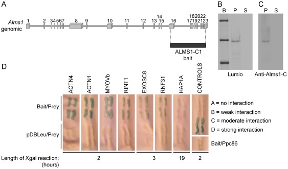
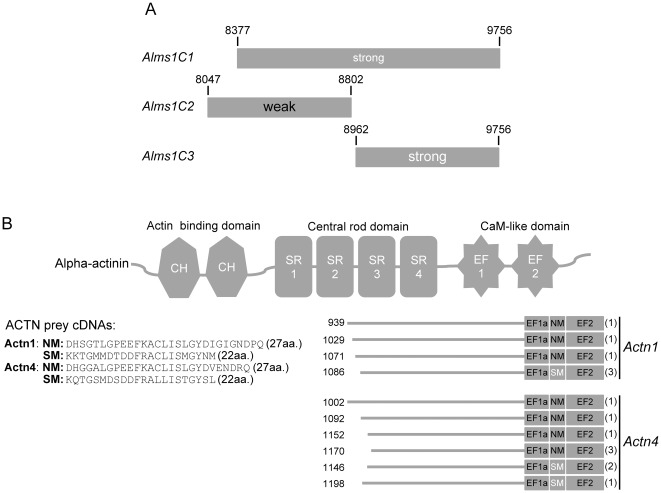
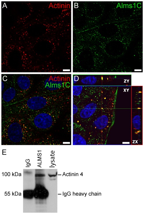
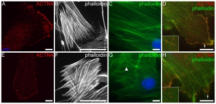
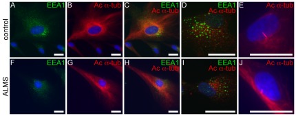
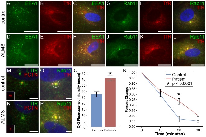
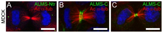
Alström syndrome (ALMS) is a progressive multi-systemic disorder characterized by cone-rod dystrophy, sensorineural hearing loss, childhood obesity, insulin resistance and cardiac, renal, and hepatic dysfunction. The gene responsible for Alström syndrome, ALMS1, is ubiquitously expressed and has multiple splice variants. The protein encoded by this gene has been implicated in ciliary function, cell cycle control, and intracellular transport. To gain better insight into the pathways through which ALMS1 functions, we carried out a yeast two hybrid (Y2H) screen in several mouse tissue libraries to identify ALMS1 interacting partners. The majority of proteins found to interact with the murine carboxy-terminal end (19/32) of ALMS1 were α-actinin isoforms. Interestingly, several of the identified ALMS1 interacting partners (α-actinin 1, α-actinin 4, myosin Vb, rad50 interacting 1 and huntingtin associated protein1A) have been previously associated with endosome recycling and/or centrosome function. We examined dermal fibroblasts from human subjects bearing a disruption in ALMS1 for defects in the endocytic pathway. Fibroblasts from these patients had a lower uptake of transferrin and reduced clearance of transferrin compared to controls. Antibodies directed against ALMS1 N- and C-terminal epitopes label centrosomes and endosomal structures at the cleavage furrow of dividing MDCK cells, respectively, suggesting isoform-specific cellular functions. Our results suggest a role for ALMS1 variants in the recycling endosome pathway and give us new insights into the pathogenesis of a subset of clinical phenotypes associated with ALMS.


Conflict of interest statement

Competing Interests: The authors have declared that no competing interests exist.

Figures

Figure 1
Figure 1. Yeast two hybrid analysis of ALMS1.
(A) Alms1-C-terminal bait used for yeast two hybrid. (B) Bacterial induction of ALMS-C lumio fusion protein reveals a 55 kDa band of expected size. B = Benchmark fluorescent ladder (Invitrogen); P = pellet; S = supernatant (C) Immunoblot using anti-ALMS-C antibody shows specificity of the bacterially expressed protein to ALMS1, as indicated by blue coloration. (D) Y2H analysis reveals protein interactors with mouse ALMS1 (carboxy-terminal end).
Figure 2
Figure 2. Y2H interaction domains of ALMS1 and α-actinin.
(A) Direct interaction tests with truncated ALMS1 constructs reveals that both constructs were sufficient for the interaction with α-actinin. However the most C-terminal construct showed the strongest interaction. (B) Alignments of α-actinin 1 & 4 prey clone sequences from all three library screens. Potential ALMS1-interacting domains include the central rod and CaM-like domains. The number of clones identified with each unique actinin prey sequence is shown in parentheses. CH = calponin homology; SR = spectrin repeat; CaM = calmodulin-like; NM-nonmuscle; SM = smooth muscle.
Figure 3
Figure 3. ALMS1 interacts with α-actinin in mammalian cells.
(A–D) Co-localization of α-actinin (A) and ALMS1-C (B) in MDCK cells. Both proteins are expressed within cytoplasmic dense bodies. Antibody overlay and orthogonal projection are shown in C & D, respectively. Scale bar = 5 µm. (E) Co-immunoprecipitation of ALMS1 and actinin from renal lysates of C57BL/6Ei mice. Lysates were incubated overnight with a C-terminal ALMS1 antibody (polyclonal rabbit) and the precipitated proteins were probed with anti-ACTN4 (polyclonal rabbit) antibody.
Figure 4
Figure 4. F-actin staining in fibroblasts.
(A,E) Staining with anti-ACTN4 (red) in control and patient fibroblasts show actinin staining along stress fibers and at focal adhesions. (B–C,E–F) F-actin stress fibers were visualized by staining with FITC-conjugated phalloidin at lower (20×: B–E) and higher (100×: C,F) magnifcations. Long densely packed fibers were observed in both samples, however, nonuniform and stunted filaments (white arrowhead) were noted in ALMS fibroblasts. Co-staining with anti-ACTN4 (red) and FITC-phalloidin (green) show actinin staining along the stress fibers (inset) and focal adhesions (white arrows) in both patient and control fibroblasts. Scale bars = 10 µm (a,c,d,e,g,h); 100 µm (b,f).
Figure 5
Figure 5. Immunostaining of microtubules and endosomes in ALMS fibroblasts.
(A–D, F–I) Patient and control fibroblasts are stained with EEA1 (green, early endosomal marker) and acetylated α-tubulin (red, microtubule markers). Panels E & J show formation of primary cilia in differentiated fibroblasts from patients and controls. Scale bars = 25 µm.
Figure 6
Figure 6. Kinetics of transferrin recycling in ALMS and control fibroblasts.
(A–L) ALMS and control fibroblasts were incubated with unlabelled transferrin for 30 minutes. TfR and nuclei are depicted in red and blue, respectively. Early and recycling endosomes were immunostained with Alexa Fluor 488-labelled EEA1 (A–F) and Rab11 (G–L), respectively. Scale bars = 25 µm. (M–N) Co-localization with pericentrin (PCTN;red) and TfR (green) shows overlay at the pericentrosome. (O–Q) Quantification of TfR and its colocalization with Rab11 at the pericentrosome. Mean fluorescence intensity per 2 µm diameter area (dashed circle) was measured by ImageJ/Fiji software and used as an estimate of the number of TfR positive endosomes. Results are from 40 patient and 47 control cells and error bars indicate ± SEM; Star indicates p<0.0001. Scale bars = 5 µm. (R) Cells were ‘pulsed’ with Tf-Alexa Fluor 647 for 30 min, followed by a ‘cold chase’ of unlabelled holo-transferrin for indicated times. Data was pooled from three fibroblast cell lines from ALMS and control subjects. The graph represents the mean +/− SEM of four independent experiments as a mean percentage of Tf internalization at each time point. The values (MFI) obtained at time 0 following the pulse were set at 100%. Star denotes p<0.0001.
Figure 7
Figure 7. Distribution of ALMS1 during cell division.
(A–C) Spatial distribution of N-terminal ALMS1 and C-terminal ALMS1 during cytokinesis. ALMS-Ntr (A, green) is found within the centrioles at the spindle poles. ALMS1-C (B–C, green) redistributes to the acto-myosin contractile ring and to the cleavage furrow during late cytokinesis. Mitotic spindles are observed by acetylated α-tubulin (red) staining. Scale bars = 5 µm.

References

    1. Marshall JD, Bronson RT, Collin GB, Nordstrom AD, Maffei P, et al. New Alstrom syndrome phenotypes based on the evaluation of 182 cases. Arch Intern Med. 2005;165:675–683. - PubMed
    1. Collin GB, Marshall JD, Ikeda A, So WV, Russell-Eggitt I, et al. Mutations in ALMS1 cause obesity, type 2 diabetes and neurosensory degeneration in Alstrom syndrome. Nat Genet. 2002;31:74–78. - PubMed
    1. Hearn T, Renforth GL, Spalluto C, Hanley NA, Piper K, et al. Mutation of ALMS1, a large gene with a tandem repeat encoding 47 amino acids, causes Alstrom syndrome. Nat Genet. 2002;31:79–83. - PubMed
    1. Marshall JD, Hinman EG, Collin GB, Beck S, Cerqueira R, et al. Spectrum of ALMS1 variants and evaluation of genotype-phenotype correlations in Alstrom syndrome. Hum Mutat. 2007;28:1114–1123. - PubMed
    1. Andersen JS, Wilkinson CJ, Mayor T, Mortensen P, Nigg EA, et al. Proteomic characterization of the human centrosome by protein correlation profiling. Nature. 2003;426:570–574. - PubMed

Publication types

MeSH terms