Comparative Study
. 2012;7(5):e38239.
doi: 10.1371/journal.pone.0038239. Epub 2012 May 31.

Comparative geno-plasticity analysis of Mycoplasma bovis HB0801 (Chinese isolate)

Affiliations
Comparative Study

Comparative geno-plasticity analysis of Mycoplasma bovis HB0801 (Chinese isolate)

Jingjing Qi et al. PLoS One. 2012.

Abstract

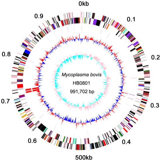
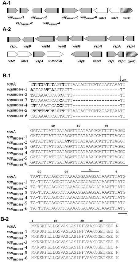
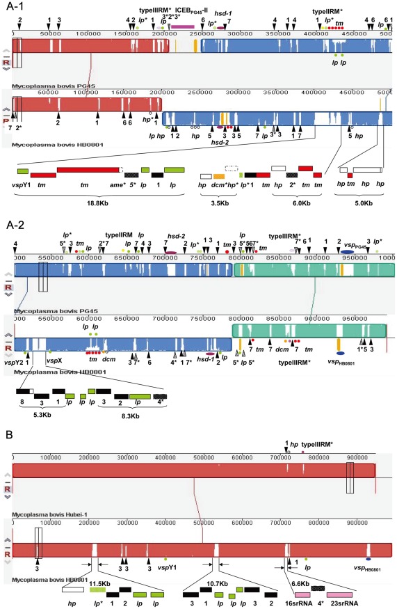
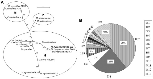
Mycoplasma bovis pneumonia in cattle has been epidemic in China since 2008. To investigate M. bovis pathogenesis, we completed genome sequencing of strain HB0801 isolated from a lesioned bovine lung from Hubei, China. The genomic plasticity was determined by comparing HB0801 with M. bovis strain ATCC® 25523™/PG45 from cow mastitis milk, Chinese strain Hubei-1 from lesioned lung tissue, and 16 other Mycoplasmas species. Compared to PG45, the genome size of HB0801 was reduced by 11.7 kb. Furthermore, a large chromosome inversion (580 kb) was confirmed in all Chinese isolates including HB0801, HB1007, a strain from cow mastitis milk, and Hubei-1. In addition, the variable surface lipoproteins (vsp) gene cluster existed in HB0801, but contained less than half of the genes, and had poor identity to that in PG45, but they had conserved structures. Further inter-strain comparisons revealed other mechanisms of gene acquisition and loss in HB0801 that primarily involved insertion sequence (IS) elements, integrative conjugative element, restriction and modification systems, and some lipoproteins and transmembrane proteins. Subsequently, PG45 and HB0801 virulence in cattle was compared. Results indicated that both strains were pathogenic to cattle. The scores of gross pathological assessment for the control group, and the PG45- and HB0801-infected groups were 3, 13 and 9, respectively. Meanwhile the scores of lung lesion for these three groups were 36, 70, and 69, respectively. In addition, immunohistochemistry detection demonstrated that both strains were similarly distributed in lungs and lymph nodes. Although PG45 showed slightly higher virulence in calves than HB0801, there was no statistical difference between the strains (P>0.05). Compared to Hubei-1, a total of 122 SNP loci were disclosed in HB0801. In conclusion, although genomic plasticity was thought to be an evolutionary advantage, it did not apparently affect virulence of M. bovis strains in cattle.


Conflict of interest statement

Competing Interests: The authors have declared that no competing interests exist.

Figures

Figure 1
Figure 1. Circular Diagram of the M. bovis HB0801 Genome Structure.
The dnaA gene is at position zero. Starting from the outside, the first circle shows the genome length (units in Mb); the second and the third circles show the locations of the predicted CDSs on the plus and minus strands, respectively, which were color-coded by COG categories (gold for translation, ribosomal structure and biogenesis; orange for RNA processing and modification; light orange for transcription; dark orange for DNA replication, recombination and repair; antique white for cell division and chromosome partitioning; pink for defense mechanisms; red for signal transduction mechanisms; peach for cell envelope biogenesis and outer membrane; deep pink for intracellular trafficking, secretion and vesicular transport; pale green for post-translational modification, protein turnover and chaperones; royal blue for energy production and conversion; blue for carbohydrate transport and metabolism; dodger blue for amino acid transport and metabolism; sky blue for nucleotide transport and metabolism; light blue for coenzyme metabolism; cyan for lipid metabolism; medium purple for inorganic ion transport and metabolism; aquamarine for secondary metabolites biosynthesis, transport and catabolism; and gray for unknown function); the fourth circle shows tRNAs (violet) and rRNAs (red); the fifth circle shows the centered GC (G+C) content of each CDS (red: above mean and blue: below mean); and the sixth circle shows the GC (G+C) skew plot.
Figure 2
Figure 2. Comparison of vsp Gene Cluster between M. bovis HB0801 and PG45.
The vsp gene loci of HB0801 (A-1) and PG45 (A-2) are shown. The locations and directions of the vsp ORFs are indicated with gray arrows. The adjacent non-vsp ORFs (ORF-1 and ORF-2) are indicated with open arrows. The locations of the putative tyrosine recombinase genes (xer C) are indicated with hatched arrows. The highly homologous regions upstream of the vsp genes are indicated with black blocks. (B) Sequence alignments for vsp upstream 5′ regions (B-1) and Vsp N-terminal regions (B-2). The HB0801 vsp gene family (vsp HB0801-1 to -6) and the vspA gene, a representative PG45 vsp gene, are compared. The numbers above the sequences indicate positions relative to the initiation codon. Nucleotides representing a putative ribosome-binding site (SD) are headlined and the initiation codon (ATG) is underlined with a horizontal arrow. The division of the vsp upstream 5′ region into two distinct cassettes is indicated by a vertical arrow at nucleotide position -72.
Figure 3
Figure 3. Genomic Comparison of M. bovis strains HB0801, PG45 and Hubei-1.
(A) Complete genome comparison between HB0801 and PG45. A-1 and A-2 represent the upstream and downstream regions of the inversion breaking sites. (B) Genome comparison between HB0801 and Hubei-1. The middle blue region of HB0801 represents the large inverted fragment compared to PG45. The numbers 1–7 represent ISMbov1-ISMbov7, respectively (black triangles); lp: putative lipoprotein gene (green dots); tm: putative transmembrane protein gene (red dots); hsd: type I restriction modification system (purple dots); dcm: DNA-methyltransferase gene (orange dots); vsp: variable surface lipoprotein (blue dots); hp: hypothetical protein gene (hollow dots). The asterisks mark pseudo genes.
Figure 4
Figure 4. Phylogenetic Analysis and Orthologous Gene Detection.
(A) Phylogenetic relationships of consensus sequences of 17 Mycoplasma strains with complete genomic sequences. The phylogenetic groups, mycoides, pneumoniae and hominis, are indicated by M, P and H, respectively. (B) Frequency of orthologous genes within the 13 genomes of different Mycoplasma species included in this analysis are listed in Table 1 (marked with *). The figures 1 to 13 on the right panel represent the number of genomes where the common orthologous genes were found. The rates in different parts of the circle represent the frequency of genes present in a single genome or shared by different genomes. The figures surrounding the circle represent orthologous gene numbers in individual parts. The 39% of genes including 887 present in a single genome represent lineage specific genes, while 10% of the genes including 224 were found in all 13 genomes, which represent the Mycoplasma core genome.
Figure 5
Figure 5. PCR Confirmation of Inversion and vsp Gene Cluster.
(A) PCR amplification specific to the upstream and downstream connection sites of the M. bovis HB0801 inversion region. Lanes 1–3 represent PCR products using the primer Inv-1 specific to upstream connection region of the inversion. Lanes 4–6 represent PCR products using primer Inv-2 specific to the downstream connection region of the inversion. Lanes 1 and 4 used the HB0801 genome with pneumonia origin as the template, Lanes 2 and 5 used the HB1007 genome with mastitis origin as the template, and Lanes 3 and 6 represent PCR products using the M. bovis ATCC® 25523™/PG45 genome with mastitis origin as the template. (B) PCR product of the vsp region in the HB0801 genome.
Figure 6
Figure 6. Immunohistochemical Staining of Lung Sections of Calves with M.bovis Infection.
The lung tissues of calves from the negative control group (A), M. bovis HB0801- (B) and PG45-infected groups (C) were detected after immunohistochemical staining with monoclonal antibody to M. bovis. The positive cells were stained brown and the negative cells were counter-stained blue with hematoxylin. The positive cells were situated in bronchiole epithelia.

Similar articles

Cited by

References

    1. Caswell JL, Archambault M. Mycoplasma bovis pneumonia in cattle. Animal Health Research Reviews. 2008;8:161–186. - PubMed
    1. Caswell JL, Bateman KG, Cai HY, Castillo-Alcala F. Mycoplasma bovis in respiratory disease of feedlot cattle. Vet Clin North Am Food Anim Pract. 2010;26:365–379. - PubMed
    1. White BJ, Hanzlicek G, Sanderson MW, Anderson DE, Larson RL. Mollicutes species and Mycoplasma bovis prevalence and association with health outcomes in beef feeder calves at arrival and initial treatment for bovine respiratory disease. Can Vet J. 2010;51:1016–1018. - PMC - PubMed
    1. Shi L. Diagnosis of Cattle Infectious Mycoplasma bovis Pneumonia. Journal of Huazhong Agricultural University. 2008;5:629–633.
    1. Wise KS, Calcutt MJ, Foecking MF, Roske K, Madupu R, et al. Complete Genome Sequence of Mycoplasma bovis Type Strain PG45 (ATCC 25523). Infection and Immunity. 2010;79:982–983. - PMC - PubMed

Publication types

Substances