Randomized Controlled Trial
. 2012 Oct;12(10):2730-43.
doi: 10.1111/j.1600-6143.2012.04144.x. Epub 2012 Jun 13.

Subclinical inflammation and chronic renal allograft injury in a randomized trial on steroid avoidance in pediatric kidney transplantation

Affiliations
Randomized Controlled Trial

Subclinical inflammation and chronic renal allograft injury in a randomized trial on steroid avoidance in pediatric kidney transplantation

M Naesens et al. Am J Transplant. 2012 Oct.

Abstract

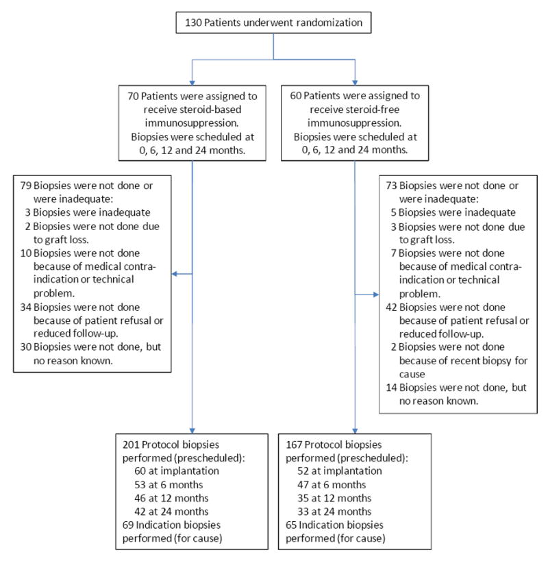
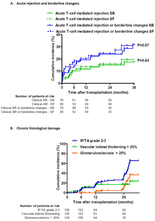
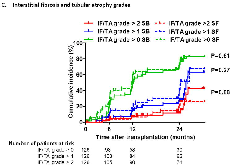
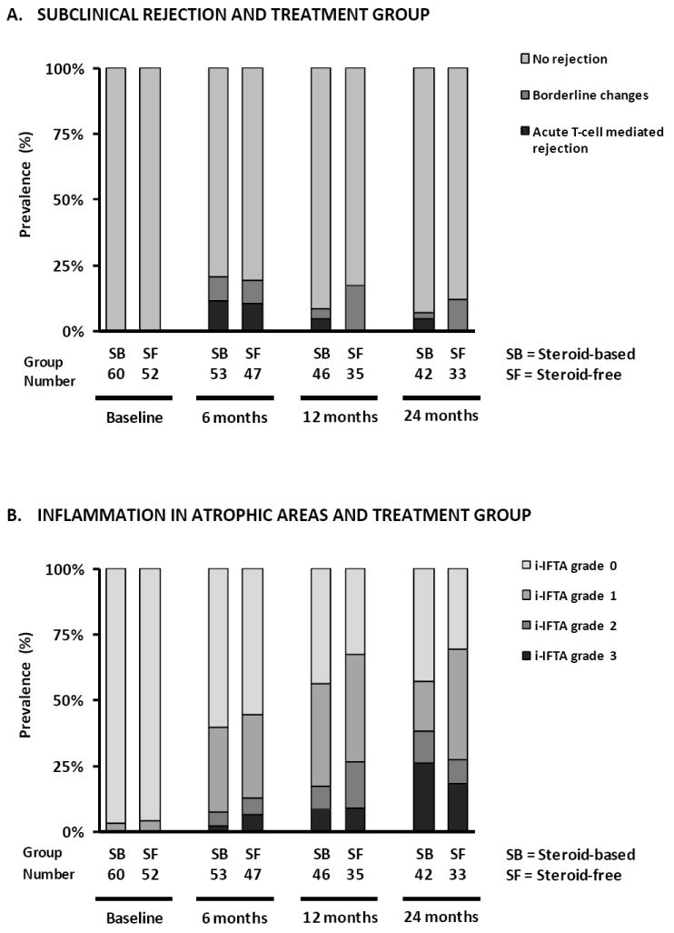
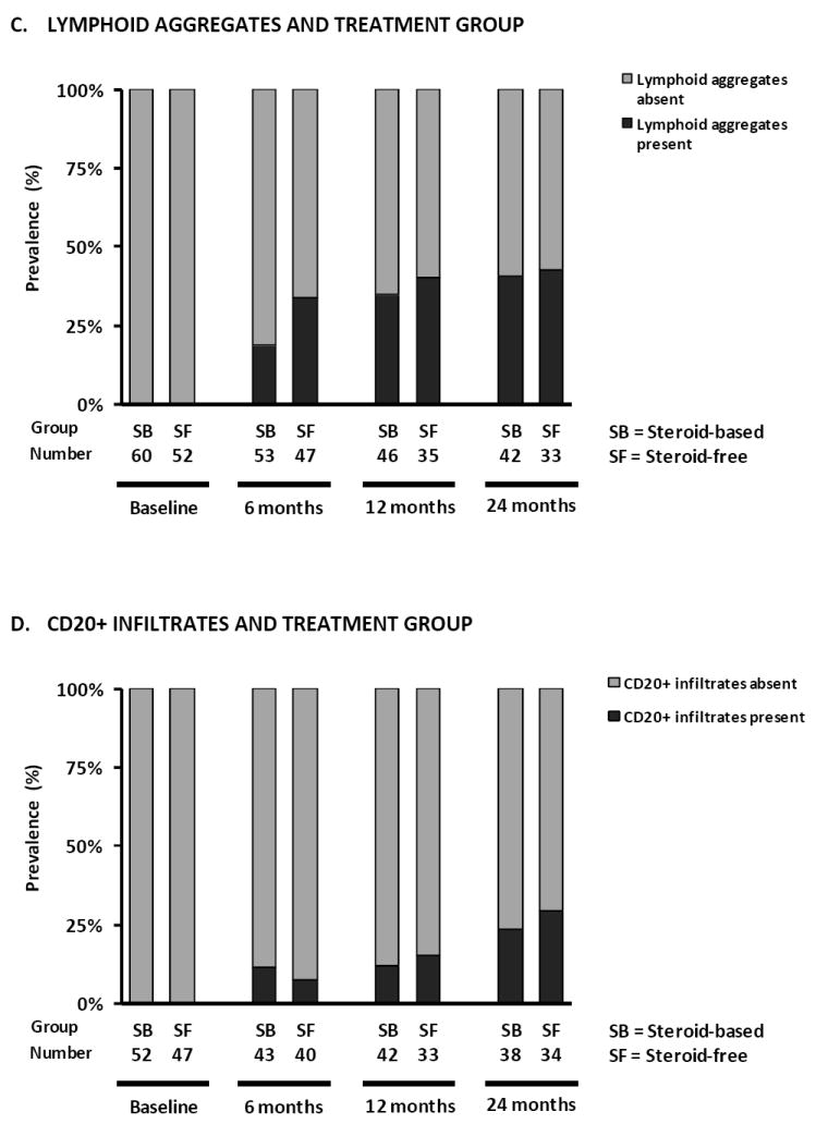
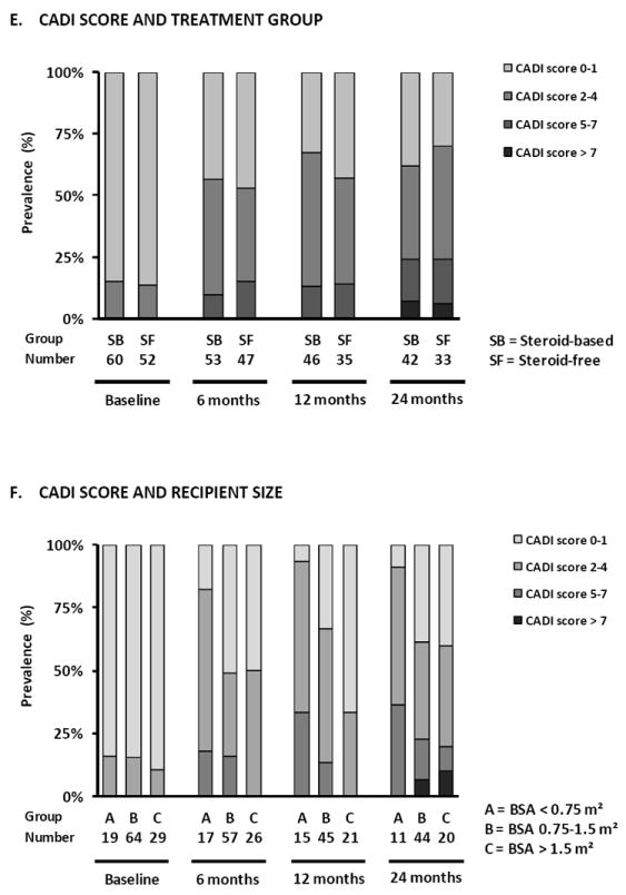
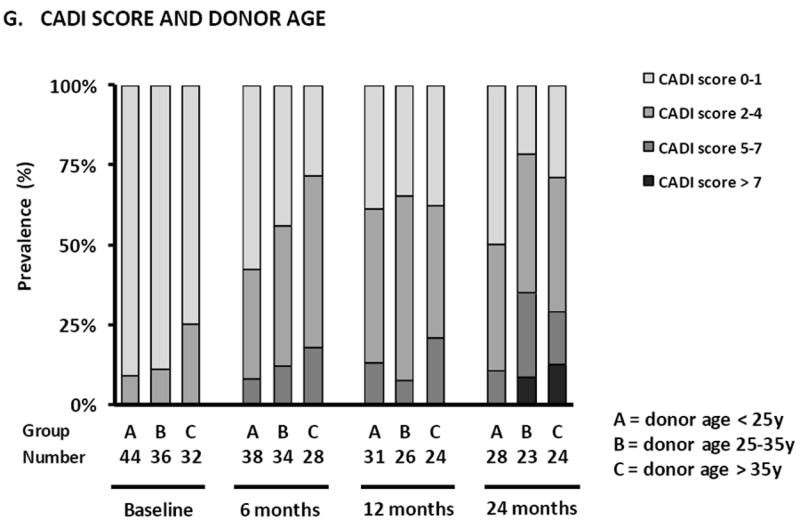
Steroid avoidance is safe and effective in children receiving kidney transplants in terms of graft function and survival, but the effects on allograft histology are unknown. In this multicenter trial, 130 pediatric renal transplant recipients were randomized to steroid-free (SF; n = 60) or steroid-based (SB; n = 70) immunosuppression, and underwent renal allograft biopsies at the time of graft dysfunction and per protocol at implantation and 6, 12 and 24 months after transplantation. Clinical follow-up was 3 years posttransplant. Subclinical acute rejection was present in 10.6% SF versus 11.3% SB biopsies at 6 months (p = 0.91), 0% SF versus 4.3% SB biopsies at 1 year (p = 0.21) and 0% versus 4.8% at 2 years (p = 0.20). Clinical acute rejection was present in 13.3% SF and 11.4% SB patients by 1 year (p = 0.74) and in 16.7% SF and 17.1% SB patients by 3 years (p = 0.94) after transplantation. The cumulative incidence of antibody-mediated rejection was 6.7% in SF and 2.9% in SB by 3 years after transplantation (p = 0.30). There was a significant increase in chronic histological damage over time (p < 0.001), without difference between SF and SB patients. Smaller recipient size and higher donor age were the main risk factors for chronic histological injury in posttransplant biopsies.

Trial registration: ClinicalTrials.gov NCT00141037.


Conflict of interest statement

DISCLOSURE

None of the authors have any conflict of interest to declare in relation to this study.

Figures

Figure 1
Figure 1. Study design and numbers of biopsies included
All patients who underwent randomization and transplantation were included in the intent-to-treat analysis.
Figure 2
Figure 2. Cumulative incidence of acute and chronic histological lesions by time after transplantation
(A) Cumulative incidence of clinical (in biopsies for cause) acute T-cell mediated rejection or borderline changes according to treatment arm. (B) Cumulative incidence of different grades of interstitial fibrosis/tubular atrophy (IFTA), vascular intimal thickening and global glomerulosclerosis by time after transplantation, expressed as 1-the Kaplan-Meier survival estimate. (C) Cumulative incidence of different grades of interstitial fibrosis/tubular atrophy (IFTA) according to treatment arm. IFTA grade 1 is IFTA encompassing <25% of the biopsy core, while grade 2 corresponds to 25-50% and grade 3 to IFTA in >50% of the biopsy. For the Kaplan–Meier estimates of event rates presented here, patients were censored at the time of their last biopsy or at 24 months if indication biopsies were performed later than 24 months. The histological lesions were scored according to the updated Banff classification on sequential biopsy specimens obtained on indication (graft dysfunction) and at prescheduled time points (protocol biopsies). P-values comparing survival distribution between SF and SB study group were obtained using the log-rank test.
Figure 3
Figure 3. Prevalence of different histological lesions at each time point after transplantation
Prevalence of subclinical rejection (A), inflammation in atrophic areas (B), lymphoid aggregates (C) and CD20+ infiltrates (D) according to treatment arm in protocol biopsies. (E) Prevalence of different degrees of CADI score according to treatment arm, (F) recipient size and (G) donor age in protocol biopsies. There were no statistically significant differences between the treatment arms in terms of the different histological lesions in GEE analysis (Supplemental Table 3). Smaller recipient size (p<0.0001) and older donor age (p<0.05) were independently associated with higher degrees of CADI score in post-transplantation biopsies in GEE analysis.

Comment in

References

    1. McDonald SP, Craig JC. Long-term survival of children with end-stage renal disease. N Engl J Med. 2004 Jun 24;350(26):2654–62. - PubMed
    1. Nankivell B, Richard J, Borrows R, Fung CL-S, O’Connell PJ, Allen R, et al. The natural history of chronic allograft nephropathy. New Engl J Med. 2003;349(24):2326–33. - PubMed
    1. El-Zoghby ZM, Stegall MD, Lager DJ, Kremers WK, Amer H, Gloor JM, et al. Identifying specific causes of kidney allograft loss. American Journal of Transplantation. 2009 Mar;9(3):527–35. - PubMed
    1. Naesens M, Kambham N, Concepcion W, Salvatierra O, Jr, Sarwal M. The evolution of non-immune histological injury and its clinical relevance in adult-sized kidney grafts in pediatric recipients. Am J Transplant. 2007;7(11):2504–14. - PubMed
    1. Chapman JR, O’Connell PJ, Nankivell BJ. Chronic renal allograft dysfunction. J Am Soc Nephrol. 2005 Oct 1;16(10):3015–26. - PubMed

Publication types

Associated data