. 2012 Jun 13:12:105.
doi: 10.1186/1471-2180-12-105.

Matrix metalloproteinase 9 contributes to gut microbe homeostasis in a model of infectious colitis

Affiliations

Matrix metalloproteinase 9 contributes to gut microbe homeostasis in a model of infectious colitis

David M Rodrigues et al. BMC Microbiol. .

Abstract

Background: Inflammatory bowel diseases are associated with increased expression of zinc-dependent Matrix Metalloproteinase 9 (MMP-9). A stark dysregulation of intestinal mucosal homeostasis has been observed in patients with chronic inflammatory bowel diseases. We therefore sought to determine the contribution of MMP-9 to the pathogenesis of Citrobacter rodentium-induced colitis and its effects on gut microbiome homeostasis.

Results: Wild-type and MMP-9-/- mice aged 5-6 weeks were challenged with C. rodentium by orogastric gavage and sacrificed either 10 or 30 days post-infection. Disease severity was assessed by histological analysis of colonic epithelial hyperplasia and by using an in vivo intestinal permeability assay. Changes in the inflammatory responses were measured by using qPCR, and the composition of the fecal microbiome evaluated with both qPCR and terminal restriction fragment length polymorphism. Activation and localization of MMP-9 to the apical surface of the colonic epithelium in response to C. rodentium infection was demonstrated by both zymography and immunocytochemistry. The pro-inflammatory response to infection, including colonic epithelial cell hyperplasia and barrier dysfunction, was similar, irrespective of genotype. Nonmetric multidimensional scaling of terminal restriction fragments revealed a different fecal microbiome composition and C. rodentium colonization pattern between genotypes, with MMP-9-/- having elevated levels of protective segmented filamentous bacteria and interleukin-17, and lower levels of C. rodentium. MMP-9-/- but not wild-type mice were also protected from reductions in fecal microbial diversity in response to the bacterial enteric infection.

Conclusions: These results demonstrate that MMP-9 expression in the colon causes alterations in the fecal microbiome and has an impact on the pathogenesis of bacterial-induced colitis in mice.


Figures
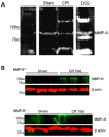
Figure 1
Figure 1
C. rodentium infection is associated with increased MMP-9 activation. (A) Representative gelatin zymogram showing the absence of MMP-9 activity in sham-infected animals and activation of MMP-9 at 10d PI with C. rodentium. Positive controls for MMP-9 were obtained from colonic homogenates from dextran sodium sulphate (DSS)-treated animals, at the peak of inflammation (8d post-DSS). (B) Representative western blot demonstrates increased activation of MMP-9 (92 kDa) in whole colon homogenates obtained from C. rodentium-infected WT and MMP-9−/− mice at 10 days PI, compared to sham-infected mice.
Figure 2
Figure 2
MMP-9−/−and WT mice infected withC. rodentiumhave similar histopathology and mucosal barrier dysfunction. (A) Representative H & E stained images of colonic tissues demonstrating C. rodentium-induced inflammation in MMP-9+/+ and MMP-9−/− mice. Scale bar, 100 μm. (B) Quantitative analysis shows significant increases in colonic crypt height in MMP-9+/+ and MMP-9−/− mice at 10d and 30d PI, compared to sham-infected mice. N = 12-18. (C) Fluorometric analysis of a 4 kDa FITC-dextran probe in serum samples obtained from WT and MMP-9−/− mice in the presence or absence of C. rodentium infection (10d and 30d PI). *P<0.05 compared to Sham WT; #P<0.05 compared to Sham MMP-9−/−. N = 7-17.
Figure 3
Figure 3
MMP-9 expression is increased withC. rodentiuminfection. Immunohistochemistry shows that MMP-9 distributed throughout the crypts (green) in uninfected WT mice is localized primarily to the apical surface of intestinal epithelium in C. rodentium-infected (10d) WT mice. Scale bar, 100 μm.
Figure 4
Figure 4
Post-infectious goblet cell hyperplasia occurs in MMP-9−/−mice. (A) Representative histology demonstrating goblet cells stained positive (red) for PAS in MMP-9+/+ and MMP-9−/− colonocytes. (B) Quantitative analysis shows similar numbers of goblet cells in WT and MMP-9−/− mice at 10d PI. A significant increase in goblet cells was observed in MMP-9−/− mice at 30d PI. *P<0.05 compared to WT-infected animals. N = 3–5.
Figure 5
Figure 5
MMP-9−/−mice demonstrate elevated baseline IL-17 transcription, compared to WT mice. Analysis of mRNA from whole-thickness distal colons obtained from infected and uninfected WT and MMP-9−/− mice for the following genes: IL-17, TNFα, IFNγ, IL-4, IL-10, FOXP3 and β–actin (housekeeping gene). *P<0.05 compared to Sham WT; #P<0.05 compared to Sham MMP-9−/−. N = 6-18.
Figure 6
Figure 6
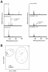
MMP-9−/−mice have an altered intestinal microbiome and decreasedC. rodentiumcolonization efficiency. (A) T-RFLP was employed to track the colonization of C. rodentium in infected mice by following the presence and intensity of the 118 bp peak on electropherograms (indicated by arrows). (B) Nonmetric multidimensional scaling of terminal restriction fragments from WT and MMP-9−/− mice reveals two distinct microbial communities. N = 15-18.
Figure 7
Figure 7
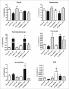
MMP-9−/−mice have a microbiome enriched in segmented filamentous bacteria. qPCR analysis of bacterial 16 s rRNA sequences specific to the following communities of bacteria: Bacillus, Bacteroides, Enterobacteriaceae, Firmicutes, Lactobacilli/Lactococci, and SFB (“A immunis”).*P<0.05 compared to Sham WT; #P<0.05 compared to Sham MMP-9−/−. N = 4-11.

Similar articles

Cited by

References

    1. Sekirov I, S.L R, Caetano L, Antunes M, Finlay B. Gut microbiota in health and disease. Physiol Rev. 2010;90:859–904. doi: 10.1152/physrev.00045.2009. - DOI - PubMed
    1. Denou E, Rezzonico E, Panoff J-M, Arigoni F, Brüssow H. A mesocosm of Lactobacillus johnsonii, Bifidobacterium longum, and Escherichia coliin the mouse gut. DNA and Cell Biology. 2009;28(8):413–422. doi: 10.1089/dna.2009.0873. - DOI - PubMed
    1. Bibiloni R, Schiffrin EJ. Intestinal host-microbe interactions under physiological and pathological conditions. Int J Inflam. 2010;2010:8. - PMC - PubMed
    1. Joossens M, Huys G, Cnockaert M, De Preter V, Verbeke K, Rutgeerts P, Vandamme P, Vermeire S. Dysbiosis of the faecal microbiota in patients with Crohn's disease and their unaffected relatives. Gut. 2011;60(5):631–637. doi: 10.1136/gut.2010.223263. - DOI - PubMed
    1. Kus JV, Gebremedhin A, Dang V, Tran S-L, Serbanescu A, Foster DB. Bile salts induce resistance to polymyxin in enterohemorrhagic Escherichia coliO157:H7. J Bacteriol. 2011;193(17):4509–4515. doi: 10.1128/JB.00200-11. - DOI - PMC - PubMed

Publication types

MeSH terms

Substances