. 2012 Jun 14;3(6):e320.
doi: 10.1038/cddis.2012.64.

A novel role for RIP1 kinase in mediating TNFα production

Affiliations

A novel role for RIP1 kinase in mediating TNFα production

D E Christofferson et al. Cell Death Dis. .

Abstract

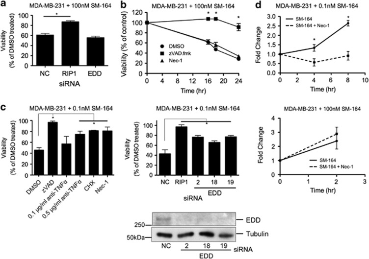
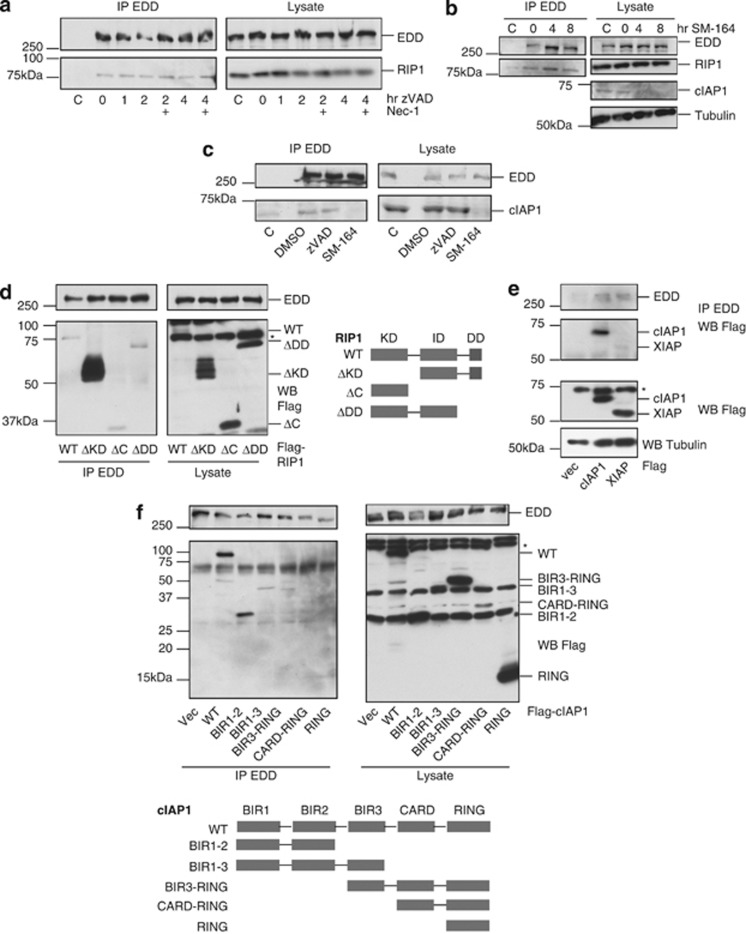
Receptor-interacting protein 1 (RIP1) is a Ser/Thr kinase with both kinase-dependent and kinase-independent roles in death receptor signaling. The kinase activity of RIP1 is required for necroptosis, a caspase-independent pathway of programmed cell death. In some cell types, the inhibition of caspases leads to autocrine production of TNFα, which then activates necroptosis. Here, we describe a novel role for RIP1 kinase in regulating TNFα production after caspase inhibition. Caspase inhibitors activate RIP1 kinase and another protein, EDD, to mediate JNK signaling, which stimulates Sp1-dependent transcription of TNFα. This pathway is independent of nuclear factor κB and also occurs after Smac mimetic/IAP antagonist treatment or the loss of TNF receptor-associated factor 2 (Traf2). These findings implicate cIAP1/2 and Traf2 as negative regulators of this RIP1 kinase-dependent TNFα production pathway and suggest a novel role for RIP1 kinase in mediating TNFα production under certain conditions.


Figures

Figure 1
Figure 1
RIP1 kinase activates TNFα production. (a) TNFα levels determined by TNFα ELISA and normalized to total protein in lysate from L929 cells treated with 20 μM zVAD.fmk ±10 μM Nec-1 for 16 h. (b) L929 or J774 cells treated with 20 μM zVAD.fmk±1.0 μg/ml anti-mTNFα or 10 μM Nec-1. Cell viability measured by ATP assay after 24 h. (c) TNFα levels in J774 cells treated with 20 μM zVAD.fmk±10 μM Nec-1 for 4 and 8 h were determined as in a. (d) TNFα levels in J774 cells treated with 20 μM zVAD.fmk±0.5 μg/ml CHX. *P<0.05, data shown are mean±S.E.M., n=3
Figure 2
Figure 2
EDD mediates TNFα production. (a) 293T cells transfected with Flag-EDD and Flag-RIP1 as indicated. Cell lysates and anti-EDD immunoprecipitates were western blotted with anti-RIP1 and anti-EDD to show both endogenous and overexpressed protein. (b) L929 cells transfected for 48 h with siRNA targeting RIP1 or one of the four different siRNAs targeting EDD were treated for 24 h with 20 μM zVAD.fmk or 10 ng/ml hTNFα and cell viability was measured by ATP assay (top) or lysates collected for western blot (bottom) to check protein knockdown. (c) J774 cells (left) or L929 cells (right) stably expressing shRNA against EDD or an empty vector control were treated with 20 μM zVAD.fmk and TNFα measured as in a. EDD knockdown shown in Figure 6e and Supplementary Figure S3a. (d) Real-time PCR of RNA isolated from J774 cells treated with zVAD.fmk with or without Nec-1 (left) or EDD stable knockdown J774 cells treated with zVAD.fmk (right). Relative levels of TNFα transcript were determined compared with GAPDH (left) or 18S rRNA (right) and the fold change was calculated by comparing with DMSO-treated cells. *P<0.05, data shown are mean±S.E.M., for all experiments n=3
Figure 3
Figure 3
Traf2 and cIAP1 inhibit RIP1- and EDD-dependent TNFα production. (a) ATP assay of L929 cells treated with 100 nℳ SM-164±TNFα-neutralizing antibody or 10 μM Nec-1 for 24 h. (b) Viability of L929 cells transfected with siRNA against RIP1, EDD, TNFR1, or negative control (NC) for 48 h and treated with 20 μM zVAD.fmk or 100 nℳ SM-164 for 24 h. (c) TNFα levels were measured by ELISA in L929 cells treated with 100 nℳ SM-164±10 μM Nec-1 (left) or stably expressing EDD shRNA or vector control (pSRP) and treated with 100 nM SM-164 (right). (d) ATP assay of L929 cells transfected with siRNA against Traf2 and treated for 72 h, with 1.0 μg/ml anti-mTNFα antibody or 10 μM Nec-1 (left). For western blot, the cells were treated with 10 μM Nec-1 from the start of transfection to block cell death and harvested after 48 h. L929 cells transfected with siRNAs targeting Traf2 and either RIP1, EDD, or TNFR1. NC±siRNA treated with zVAD.fmk used as positive control. Viability was calculated as the ratio of the luminescence values of Traf2+siRNA to untreated NC+siRNA (right). *P<0.05, data shown are mean±S.E.M., for all experiments n=3
Figure 4
Figure 4
Smac mimetic induces RIP1 kinase- and EDD-dependent TNFα production in MDA-MB-231 cells. (a) ATP assay of MDA-MB-231 cells transfected with NC, RIP1, or EDD siRNA for 48 h and treated with 100 nℳ SM-164 for 24 h. (b) Time course of MDA-MB-231 cells treated with 100 nℳ SM-164±20 μM zVAD.fmk or 10 μM Nec-1. Cell viability was measured by ATP assay. (c) ATP assay of MDA-MB-231 cells treated with 0.1 nℳ SM-164±20 μM zVAD.fmk, anti-hTNFα-neutralizing antibody, 10 μg/ml CHX, or 10 μM Nec-1 for 24 h (left). MDA-MB-231 cells transfected with the indicated siRNA for 48 h and treated with 0.1 nℳ SM-164 for 24 h or harvested for western blot (right). (d) RT-PCR of RNA from MDA-MB-231 cells treated with 0.1 nℳ SM-164 (top) or 100 nℳ SM-164 (bottom)±10 μM Nec-1 for 2, 4, or 8 h as indicated. Relative levels of TNFα transcript determined by normalizing to 18S rRNA transcript and fold change calculated in comparison with DMSO-treated cells. *P<0.05, data shown are mean±S.E.M., for all experiments n=3, western blots were repeated two times with similar results
Figure 5
Figure 5
Complex of EDD, RIP1, and cIAP1 activate TNFα production. (a) J774 cells treated with 20 μM zVAD.fmk ±10 μM Nec-1 and immunoprecipitated with anti-EDD or an isotype-matched control antibody (C). The immunoprecipitated proteins were western blotted with anti-RIP1 (representative of three independent experiments). (b) L929 cells were treated with 100 nℳ SM-164 and EDD immunoprecipitated from cells and western blotted for RIP1. (c) L929 cells treated with 20 μM zVAD.fmk or 100 nℳ SM-164 for 7 h and EDD immunoprecipitated and western blotted for cIAP1 interaction. (d) EDD immunoprecipitated from 293T cells expressing EDD and the indicated Flag-RIP1 construct. IP was western blotted with anti-Flag to detect RIP1 interaction. (e) 293T cells expressing EDD and Flag-cIAP1, Flag-XIAP, or vector control. EDD immunoprecipitated and western blotted with anti-Flag to detect IAP binding. (f) EDD immunoprecipitated from 293T cells expressing indicated Flag-cIAP1 construct and western blotted with anti-Flag to detect interaction of cIAP1 construct. All experiments were repeated at least two times. * Indicates a non-specific band
Figure 6
Figure 6
RIP1 and EDD activate JNK signaling and TNFα production. (a) L929 cells treated with 20 μM zVAD.fmk or 100 nℳ SM-164 ±10 μM Nec-1 for 2, 4, and 8 h. Cells lysates were western blotted to determine NF-κB activation (representative of two independent experiments). (b) WT J774 cells (top) or stable EDD knockdown cells (bottom) treated with 1 μg/ml LPS±Nec-1 for 2 h and TNFα levels measured by ELISA in the cell lysate (NS). (c) ATP assay of L929 cells treated as indicated±10 μM Nec-1 or 1 μg/ml JNK inhibitor SP600125 for 24 h. (d) J774 TNFα mRNA levels determined by RT-PCR after treatment with 20 μM zVAD.fmk ±1 μg/ml SP600125 for 2 and 4 h. Fold change calculated by comparing with GAPDH control and normalizing to untreated cells. (e) J774 cells (left) or J774 EDD stable knockdown cell lines (right) were treated with zVAD.fmk±Nec-1 as indicated for 15 min, 30 min, 1, 2, or 4 h and western blotted with anti-phospho-JNK (pJNK) (representative of three independent experiments).(f) ATP assay of L929 cells transfected with siRNA targeting Sp1 for 48 h and treated with 20 μM zVAD.fmk, 100 nℳ SM-164, or 10 ng/ml hTNFα for 24 h. Cell lysates western blotted to confirm knockdown of Sp1. *P<0.05, data shown are mean±S.E.M., for all experiments n=3
Figure 7
Figure 7
Model of RIP1- and EDD-mediated pathway of TNFα production. RIP1 kinase-dependent TNFα production can be activated by the treatment with zVAD.fmk, SM to degrade cIAP1/2, or by knockdown of Traf2. EDD, RIP1, and cIAP1 are constitutively bound and RIP1 kinase and EDD activate JNK signaling after stimulation. Traf2 and cIAP1 also constitutively interact. JNK activates the transcription factors AP-1 and Sp1, which transcribe TNFα mRNA. Nec-1 inhibits the activation of JNK and production of TNFα. TNFα produced can elicit inflammatory responses and induce cell death in the surrounding cells

References

    1. Holler N, Zaru R, Micheau O, Thome M, Attinger A, Valitutti S, et al. Fas triggers an alternative, caspase-8-independent cell death pathway using the kinase RIP as effector molecule. Nat Immunol. 2000;1:489–495. - PubMed
    1. Degterev A, Hitomi J, Germscheid M, Ch'en IL, Korkina O, Teng X, et al. Identification of RIP1 kinase as a specific cellular target of necrostatins. Nat Chem Biol. 2008;4:313–321. - PMC - PubMed
    1. Degterev A, Huang Z, Boyce M, Li Y, Jagtap P, Mizushima N, et al. Chemical inhibitor of nonapoptotic cell death with therapeutic potential for ischemic brain injury. Nat Chem Biol. 2005;1:112–119. - PubMed
    1. Bertrand MJ, Milutinovic S, Dickson KM, Ho WC, Boudreault A, Durkin J, et al. cIAP1 and cIAP2 facilitate cancer cell survival by functioning as E3 ligases that promote RIP1 ubiquitination. Mol Cell. 2008;30:689–700. - PubMed
    1. Park SM, Yoon JB, Lee TH. Receptor interacting protein is ubiquitinated by cellular inhibitor of apoptosis proteins (c-IAP1 and c-IAP2) in vitro. FEBS Lett. 2004;566:151–156. - PubMed

Publication types

LinkOut - more resources