. 2012 Sep;82(3):488-99.
doi: 10.1124/mol.112.078295. Epub 2012 Jun 13.

Casein kinase 2α regulates multidrug resistance-associated protein 1 function via phosphorylation of Thr249

Affiliations

Casein kinase 2α regulates multidrug resistance-associated protein 1 function via phosphorylation of Thr249

Elzbieta I Stolarczyk et al. Mol Pharmacol. 2012 Sep.

Abstract

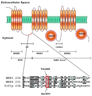
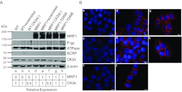
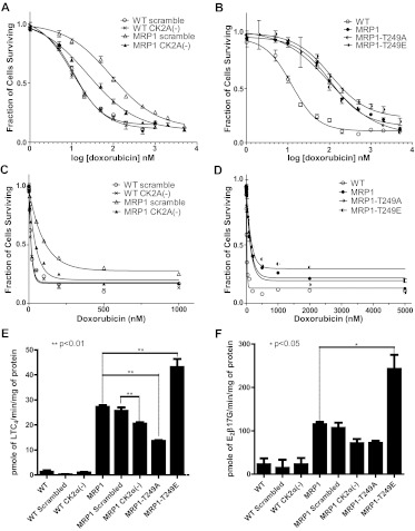
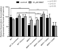
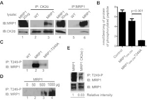
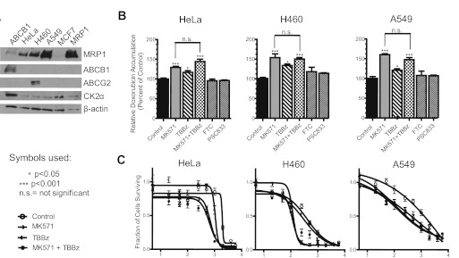
We have shown previously that the function of Ycf1p, yeast ortholog of multidrug resistance-associated protein 1 (MRP1), is regulated by yeast casein kinase 2α (Cka1p) via phosphorylation at Ser251. In this study, we explored whether casein kinase 2α (CK2α), the human homolog of Cka1p, regulates MRP1 by phosphorylation at the semiconserved site Thr249. Knockdown of CK2α in MCF7-derived cells expressing MRP1 [MRP1 CK2α(-)] resulted in increased doxorubicin sensitivity. MRP1-dependent transport of leukotriene C(4) and estradiol-17β-d-glucuronide into vesicles derived from MRP1 CK2α(-) cells was decreased compared with MRP1 vesicles. Moreover, mutation of Thr249 to alanine (MRP1-T249A) also resulted in decreased MRP1-dependent transport, whereas a phosphomimicking mutation (MRP1-T249E) led to dramatic increase in MRP1-dependent transport. Studies in tissue culture confirmed these findings, showing increased intracellular doxorubicin accumulation in MRP1 CK2α(-) and MRP1-T249A cells compared with MRP1 cells. Inhibition of CK2 kinase by 2-dimethylamino-4,5,6,7-tetrabromo-1H-benzimidazole resulted in increased doxorubicin accumulation in MRP1 cells, but not in MRP1 CK2α(-), MRP1-T249A, or MRP1-T249E cells, suggesting that CK2α regulates MRP1 function via phosphorylation of Thr249. Indeed, CK2α and MRP1 interact physically, and recombinant CK2 phosphorylates MRP1-derived peptide in vitro in a Thr249-dependent manner, whereas knockdown of CK2α results in decreased phosphorylation at MRP1-Thr249. The role of CK2 in regulating MRP1 was confirmed in other cancer cell lines where CK2 inhibition decreased MRP1-mediated efflux of doxorubicin and increased doxorubicin cytotoxicity. This study supports a model in which CK2α potentiates MRP1 function via direct phosphorylation of Thr249.


Figures

Fig. 1.
Fig. 1.
A putative CK2 phosphorylation site is semiconserved in human MRP1. The structure of MRP1 consists of a unique for ABCC subfamily NTE and a common ABC “core” region. The NTE is composed of one membrane-spanning domain (MSD0) and a cytosolic linker (L0), whereas the ABC “core” region, common to all ABC proteins, contains two membrane-spanning domains (MSD1 and MSD2) and two nucleotide-binding domains (NBD1 and NBD2). The NTE is thought to be involved in protein trafficking, stability, substrate binding, transport (Bakos et al., 2000; Fernández et al., 2002; Mason and Michaelis, 2002), and possibly in protein dimerization (Doyle et al., 1998; Yang et al., 2007, 2010). The full-length ABCC protein sequences, MRP1, MRP6, and Ycf1p were aligned using ClustalW. Alignment revealed that Ser251 of NTE, recently characterized as Cka1p regulatory site on Ycf1p (Paumi et al., 2008; Pickin et al., 2010) is semiconserved in human MRP1 as Thr249. The site is also conserved in murine MRP6 and was shown to be phosphorylated in vivo (Villén et al., 2007).
Fig. 2.
Fig. 2.
Expression of relevant proteins in MCF7-derived cell lines used in this study. Cell lines depicted in A and B are designated as follows: a, WT cells. b, WT scrambled. c, WT CK2α(−). d, MRP1. e, MRP1 scrambled. f, MRP1 CK2α(−). g, MRP1-T249A. h, MRP1-T249E. A, cell lysates (100 μg) were subjected to electrophoresis on 9% SDS-PAGE gel and immunoblotted with antibodies specific for human MRP1 (mouse monoclonal QCRL-1, 1:200; Santa Cruz Biotechnology), Na+/K+-ATPase α-1 (mouse monoclonal Na+/K+ ATPase, 1:20,000; Millipore Corp.), ABCB1 (mouse C219, 1 μg/ml; Calbiochem), ABCG2 (BXP-21; Santa Cruz Biotechnology), CK2α [goat CK2α (C-18), 1:500; Santa Cruz Biotechnology], β-actin (mouse β-actin, 1:1000; Abcam Inc., Cambridge, MA). Densitometric analysis was performed with use of Adobe Photoshop CS4, and expression of MRP1 and CK2α was normalized to β-actin. B, immunofluorescence staining of all MCF7-derived cell lines showing no visible changes in cellular localization of overexpressed MRP1 protein compared with control (MRP1): red, MRP1 signal (MRPr1 antibody, Alexa 568); blue, 4,6-diamidino-2-phenylindole nuclear stain.
Fig. 3.
Fig. 3.
Knockdown of CK2α and MRP1-Thr249 mutations alter MRP1 function in tissue culture and in vitro transport assays. Doxorubicin cytotoxicity profiles were determined by MTT assay as described under Materials and Methods. Points represent mean values ± 95% confidence intervals of three or more separate experiments performed with repeated measures in 96-well plates. The data were normalized to the baseline absorbance. Four-parameter logistic nonlinear regression model fit and fraction of cell survival-doxorubicin concentration data plots were generated with use of GraphPad Prism 5. Summary statistics derived from these data are listed in Table 1. A, four-parameter logistic nonlinear regression curve fit for WT scrambled, WT CK2α (−), MRP1 scrambled, MRP1 CK2α(−). B, four-parameter logistic nonlinear regression curve fit for WT, MRP1, MRP1-T249A, and MRP1-T249E. C, fraction of cell survival-doxorubicin concentration data plot for WT scrambled, WT CK2α (−), MRP1 scrambled, and MRP1 CK2α(−). D, fraction of cell survival-doxorubicin concentration data plot for WT, MRP1, MRP1-T249A, and MRP1-T249E. In vitro transport into inside-out vesicles prepared from presented cell lines was measured for two 3H-labeled MRP1 substrates, LTC4 (E) and E217βG (F). Transport experiments were performed in triplicate or greater, and the bar graph shows mean values ± S.D. Statistical analysis was performed using one-way ANOVA followed by Bonferroni post test.
Fig. 4.
Fig. 4.
CK2α knockdown, inhibition of CK2, and mutation of the putative CK2 phosphorylation site at Thr249 to alanine results in decreased MRP1-mediated doxorubicin efflux. Doxorubicin accumulation assays were performed as described under Materials and Methods. Where indicated, cells were pretreated with 10 μM DMAT, a potent CK2α inhibitor, before the addition of doxorubicin (gray bars). The bars represent mean value ± S.D. of three or more separate experiments with treatments performed in triplicate or greater. Statistical analysis was performed using ANOVA followed by Bonferroni post test.
Fig. 5.
Fig. 5.
A, MRP1 and CK2α proteins interact physically. Protein lysates prepared from WT and MRP1 cells were subjected to CoIP with an antibody against CK2α (goat CK2α; lanes 2 and 3) or an antibody against MRP1 (mouse QCRL-1; lanes 5 and 6). Protein A/G PLUS Agarose was added to precipitate the immunocomplex and proteins were analyzed by immunoblotting (IB). Small aliquot of MRP1 lysate was loaded as a positive control for immunoblotting (lanes 1 and 4). Reactions were carried-out in triplicate and blots shown here are representative of the series. B, phosphorylation of MRP1-Thr249 consensus site by recombinant CK2α is dependent on Thr249. The ability of recombinant human CK2α to phosphorylate MRP1-Thr249 consensus site was assessed by in vitro kinase assay as described under Materials and Methods. Synthetic peptides biotin-Ahx-RRRADDSDDDDDK (Control), biotin-Ahx- LNKEDTSEQVV (MRP1244–255), and biotin-Ahx- LNKEDASEQVV (MRP1244- 254T249A) were incubated in the presence of recombinant human CK2α and 32P-labeled ATP. All experiments were normalized to a zero time point and performed in triplicate, and results are reported as nanomoles of phosphorylated peptide formed per 5 min of reaction per milligram of recombinant protein. Statistical analysis was performed using Student's t test (control peptide served as a positive control to test for activity of the recombinant CK2α protein and therefore was not taken into consideration for statistical analysis). C, MRP1-Thr249-P antibody pulls down MRP1 protein from MRP1 cells but not from MRP1-T249A line. Custom-made rabbit antibody against synthetic peptide corresponding to MRP1-Thr249-P consensus site [WSLNKEDT(p)SEQVVP] was used to immunoprecipitate (IP) MRP1 protein from membrane fractions prepared from WT, MRP1, and MRP1-T249A cells, followed by IB with MRP1 antibody (QCRL-1). D, test of linearity carried out by immunoprecipitation with fixed amount of MRP1-Thr249-P antibody on increasing amount of MRP1 membrane fraction. E, knockdown of CK2α results in decreased MRP1 phosphorylation at Thr249. Membrane fractions prepared from MRP1 and MRP1 CK2α(−) cells were subjected to immunoprecipitation with rabbit MRP1-Thr249-P antibody (top). Relative phosphorylation (phosphorylated MRP1/ total MRP1) was determined by densitometric analysis with use of Adobe Photoshop CS4.
Fig. 6.
Fig. 6.
Inhibition of CK2 in HeLa, H460, and A549 cancer cell lines results in decreased MRP1-dependent doxorubicin efflux. A, Western blot showing expression of MRP1, ABCB1, ABCGw2, and CK2α proteins in HeLa, H460, A549, and MCF7 (WT) and MCF7/MRP1 (MRP1) cell lines. In lane 1, ABCB1-expressing cell line was loaded as a positive control for immunoblotting of ABCB1. B, doxorubicin accumulation assays were performed as described under Materials and Methods. Where indicated, cells were pretreated with 50 μM MK571, 40 μM TBBz, 10 μM FTC, or 1 μM PSC883 for 1 h before the addition of doxorubicin. Experiments were performed in triplicate, and results were presented as mean values ± S.D. Statistical analysis was performed using one-way ANOVA followed by Bonferroni post-test. C, cytotoxicity profiles of doxorubicin were determined by MTT assay as described under Materials and Methods. Points represent mean values ± 95% confidence intervals of two or three separate experiments performed with repeated measures in 96-well plates. The data were normalized to the baseline absorbance, and four-parameter logistic nonlinear regression curves were generated with use of GraphPad Prism 5. Summary statistics derived from these data are listed in Table 2.

Similar articles

Cited by

References

    1. Ausubel FM, Brent R, Kingston RE, Moore FF, Seidman JG, Smith JA, Struhl K. (1987) Current Protocols in Molecular Biology, Greene Publishing Associates/Wiley Interscience, New York
    1. Bakos E, Evers R, Calenda G, Tusnády GE, Szakács G, Váradi A, Sarkadi B. (2000) Characterization of the amino-terminal regions in the human multidrug resistance protein (MRP1). J Cell Sci 113:4451–4461 - PubMed
    1. Burnett G, Kennedy EP. (1954) The enzymatic phosphorylation of proteins. J Biol Chem 211:969–980 - PubMed
    1. Campling BG, Pym J, Baker HM, Cole SP, Lam YM. (1991) Chemosensitivity testing of small cell lung cancer using the MTT assay. Br J Cancer 63:75–83 - PMC - PubMed
    1. Campling BG, Pym J, Galbraith PR, Cole SP. (1988) Use of the MTT assay for rapid determination of chemosensitivity of human leukemic blast cells. Leuk Res 12:823–831 - PubMed

Publication types

MeSH terms