. 2012 Aug;3(4):373-86.
doi: 10.1007/s13244-011-0142-z. Epub 2012 Jan 15.

MRI of the lung (3/3)-current applications and future perspectives

Affiliations

MRI of the lung (3/3)-current applications and future perspectives

Jürgen Biederer et al. Insights Imaging. 2012 Aug.

Abstract

Background: MRI of the lung is recommended in a number of clinical indications. Having a non-radiation alternative is particularly attractive in children and young subjects, or pregnant women.

Methods: Provided there is sufficient expertise, magnetic resonance imaging (MRI) may be considered as the preferential modality in specific clinical conditions such as cystic fibrosis and acute pulmonary embolism, since additional functional information on respiratory mechanics and regional lung perfusion is provided. In other cases, such as tumours and pneumonia in children, lung MRI may be considered an alternative or adjunct to other modalities with at least similar diagnostic value.

Results: In interstitial lung disease, the clinical utility of MRI remains to be proven, but it could provide additional information that will be beneficial in research, or at some stage in clinical practice. Customised protocols for chest imaging combine fast breath-hold acquisitions from a "buffet" of sequences. Having introduced details of imaging protocols in previous articles, the aim of this manuscript is to discuss the advantages and limitations of lung MRI in current clinical practice.

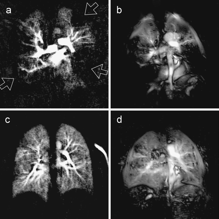
Conclusion: New developments and future perspectives such as motion-compensated imaging with self-navigated sequences or fast Fourier decomposition MRI for non-contrast enhanced ventilation- and perfusion-weighted imaging of the lung are discussed. Main Messages • MRI evolves as a third lung imaging modality, combining morphological and functional information. • It may be considered first choice in cystic fibrosis and pulmonary embolism of young and pregnant patients. • In other cases (tumours, pneumonia in children), it is an alternative or adjunct to X-ray and CT. • In interstitial lung disease, it serves for research, but the clinical value remains to be proven. • New users are advised to make themselves familiar with the particular advantages and limitations.


Figures

Fig. 1
Fig. 1
A 29-year-old female with cystic fibrosis. The axial T2-weighted (BLADE; a) and the volumetric contrast-enhanced 3D-GRE (VIBE; b) breath-hold acquisitions show severe bronchiectasis, bronchial wall thickening, mucus plugging, pleural effusion as well as a destructed middle lobe. The perfusion subtraction image (c) shows a severely impaired perfusion pattern with loss of perfusion in several areas. The maximum enhancement (MAX) and time to peak anhancement (TTP maps) allow for a further characterisation of the perfusion impairment. Most areas with impaired perfusion show a reduced (MAX map) and delaid (TTP map) perfusion. Notice the area in the left upper lobe with reduced but not delayed perfusion (arrowhead)
Fig. 2
Fig. 2
A 55-year-old patient with acute pulmonary embolism. Coronal steady-state free precession images acquired during free breathing (a) and contrast-enhanced coronal 3d flash angiogram acquired in breathhold (b; embolus inside the right lower lobe artery circled); c series of subtracted images from the first pass perfusion study, perfusion deficits marked with open arrows at the image obtained at peak lung enhancement; 1.5-T MRI scanner
Fig. 3
Fig. 3
A 56-year-old female patient with small cell lung cancer. The transverse T2-weighted fat-saturated (a) and T1-weighted contrast-enhanced fat-saturated 3D-GRE images (b, c) show a large, centrally necrotic mass in the left upper lobe with large peri-hilar lymph node metastases. Note the high soft tissue contrast between alelectatic lung (open arrow), small rim of solid tumour (filled arrow) and colliquated central portion of the mass (asterisk)
Fig. 4
Fig. 4
A 13-year-old girl with suspected organising pneumonia (BOOP) in both lungs. Transverse T2-weighted TSE images (a) were acquired with the navigator technique (sample volume placed on the dome of the right liver lobe). The open arrow indicates an oval-shaped consolidation with pleural contact in the lower left lobe and moderate signal intensity. Coronal contrast-enhanced fat-saturated T1-weighted GRE images (b) were acquired with the breathhold technique. The open arrow indicates the oval-shaped consolidation in the lower left lobe with contrast enhancement; this is interpreted as an indicator of an active inflammatory process
Fig. 5
Fig. 5
Infiltrative disorder of the lung. Typical fibrotic changes in a patient with interstitial pulmonary fibrosis. Extensive reticulation and architectural distortion predominant in the subpleural regions of the lung are well demonstrated by the axial (a, b) and coronal (ce) MR images obtained using the half-Fourier single-shot fast spin echo (a, c, e) and post-constrast volume interpolated T1-weighted GRE (b, e) sequences
Fig. 6
Fig. 6
Subtle subpleural reticulation in a patient with fibrotic-predominant NSIP. The interlobular reticulation (thin arrows) is more evident after contrast administration (c and f). A perfusion defect (arrowhead in e) is associated to the peripheral fibrotic changes at the left lateral costo-phrenic angle (arrowheads in d and f)
Fig. 7
Fig. 7
Fibrosis associated with rounded consolidation. a) Subpleural reticular changes are visualised at the periphery of the lungs (thin arrows). b) After contrast administration the subtle linear enhancement at the pulmonary-chest wall interface indicates abnormal findings related to subpleural fibrosis (thin arrows). A rounded consolidation is present on the left in the lingula suspected for lung tumour in ILD (asterisk)
Fig. 8
Fig. 8
Bilateral hilar and mediastinal adenomegalies in sarcoidosis. Node enlargement (arrows) is demonstrated with gradient echo images before (a) and after administration of contrast material (b). Coronal perfusion images indicate vascular compression at the right hilum (arrow, c) and a wedge-shaped perfusion defect (asterisk, d)
Fig. 9
Fig. 9
Twenty-three year-old female with acute pulmonary embolism at the time point of diagnosis (a, b) and at follow-up study after 6 months (c, d). The initial dynamic contrast enhanced (DCE) study (a) as well as the perfusion-weighted Fourier-decomposition (FD) image (b) demonstrate multiple perfusion defects (open arrows). In the follow up study, both techniques (DCE; c and FD; d) demonstrate an almost homogeneous lung perfusion after effective anticoagulation

References

    1. Puderbach M, Hintze C, Ley S, Eichinger M, Kauczor H-U, Biederer J. MR imaging of the chest: a practical approach at 1.5T. Eur J Radiol. 2007;64:345–355. doi: 10.1016/j.ejrad.2007.08.009. - DOI - PubMed
    1. Biederer J, Hintze C, Fabel M, Jakob PM, Horger W, Graessner J, Bolster BD, Heller M. MRI of the lung—ready…get set…go! Magnetom Flash. 2011;46:6–15.
    1. Biederer J, Both M, Graessner J, Liess C, Jakob P, Reuter M, Heller M. Lung morphology: fast MR imaging assessment with a volumetric interpolated breath-hold technique: initial experience with patients. Radiology. 2003;226:242–249. doi: 10.1148/radiol.2261011974. - DOI - PubMed
    1. Biederer J, Reuter M, Both M, Muhle C, Grimm J, Graessner J, Heller M. Analysis of artefacts and detail resolution of lung MRI with breath-hold T1-weighted gradient-echo and T2-weighted fast spin-echo sequences with respiratory triggering. Eur Radiol. 2002;12:378–384. doi: 10.1007/s00330-001-1147-7. - DOI - PubMed
    1. Biederer J, Hintze C, Fabel M. MRI of pulmonary nodules: technique and diagnostic value. Cancer Imaging. 2008;8:125–130. doi: 10.1102/1470-7330.2008.0018. - DOI - PMC - PubMed

LinkOut - more resources