. 2012 Aug;3(4):355-71.
doi: 10.1007/s13244-011-0146-8. Epub 2012 Feb 13.

MRI of the lung (2/3). Why … when … how?

Affiliations

MRI of the lung (2/3). Why … when … how?

J Biederer et al. Insights Imaging. 2012 Aug.

Abstract

Background: Among the modalities for lung imaging, proton magnetic resonance imaging (MRI) has been the latest to be introduced into clinical practice. Its value to replace X-ray and computed tomography (CT) when radiation exposure or iodinated contrast material is contra-indicated is well acknowledged: i.e. for paediatric patients and pregnant women or for scientific use. One of the reasons why MRI of the lung is still rarely used, except in a few centres, is the lack of consistent protocols customised to clinical needs.

Methods: This article makes non-vendor-specific protocol suggestions for general use with state-of-the-art MRI scanners, based on the available literature and a consensus discussion within a panel of experts experienced in lung MRI.

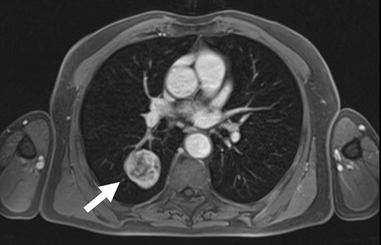
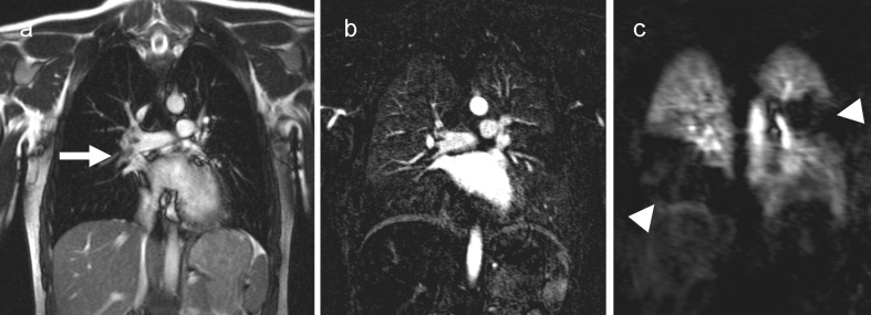
Results: Various sequences have been successfully tested within scientific or clinical environments. MRI of the lung with appropriate combinations of these sequences comprises morphological and functional imaging aspects in a single examination. It serves in difficult clinical problems encountered in daily routine, such as assessment of the mediastinum and chest wall, and even might challenge molecular imaging techniques in the near future.

Conclusion: This article helps new users to implement appropriate protocols on their own MRI platforms. Main Messages • MRI of the lung can be readily performed on state-of-the-art 1.5-T MRI scanners. • Protocol suggestions based on the available literature facilitate its use for routine • MRI offers solutions for complicated thoracic masses with atelectasis and chest wall invasion. • MRI is an option for paediatrics and science when CT is contra-indicated.


Figures

Fig. 1
Fig. 1
A 66-year-old male patient with fever and coughing, clinically suspected pneumonia. The plain chest X-ray (a) demonstrates a dense infiltrate in the left lower lung lobe which is confirmed on non-contrast-enhanced low dose CT (b; arrows). The patient volunteered to undergo MRI on the same day. Multi-breath-hold coronal T2-weighted fast spin echo (c) and single breath-hold T1-weighted 3D GRE imaging (d) as well as free breathing coronal steady state SSFP (e) and multi-breath-hold fat-saturated T2-weighted fast spin echo series (f) clearly demonstrate the infiltrates with particularly high signal on T2-weighted images
Fig. 2
Fig. 2
A 64-year-old woman with the incidental finding of an unspecific, 4-mm nodule in the right middle lobe. The nodule (open arrow) is clearly depicted on the coronal multi-breath-hold T2-weighted (a) and transverse contrast-enhanced, fat-saturated 3D GRE images (b)
Fig. 3
Fig. 3
Diffusion-weighted imaging highlighting pleural metastases of hepatocellular carcinoma at the right diaphragm (left, open arrow). The same spots are only hardly visible on the post-contrast fat-saturated breath-hold T1-weighted 3D GRE sequence (right).
Fig. 4
Fig. 4
An 18-year-old male cystic fibrosis patient, coronal T2-weighted half Fourier fast spin echo sequence (a) and coronal subtraction perfusion image (b). Notice the severe mucus plugging in the morphological T2-weighted image. The subtraction perfusion image shows correspoding areas with perfusion loss due to hypoxic vasoconstriction. Due to redistribution of perfusion both lower lobes show a high perfusion signal
Fig. 5
Fig. 5
Lung MRI of a 37-year-old male patient with cystic fibrosis. Coronal T2-weighted, respiration triggered and transverse breath-hold T1-weighted 3D GRE images show peripheral airways with enhanced signal due to mucus plugging. Note the “tree-in-bud” sign similar to the typical appearance on CT (dashed circles)
Fig. 6
Fig. 6
A 6-year-old child with lung metastases of osteosarcoma. Both acquisitions, the free breathing steady state free precession series (a) and the respiration triggered (navigator triggered) series (b) show a large mass with high signal intensity in the right upper lung lobe in expiration
Fig. 7
Fig. 7
Pneumonia (asterisk), chambered pleural effusion (arrowheads) and abscess (arrow) in the right lower chest of a 6-year-old child, images acquired in T2-weighted triggered fast spin echo technique
Fig. 8
Fig. 8
Recent fracture of the left 5th rib as incidental finding in a 29-year-old female volunteer with left chest pain, hardly visible on the non-contrast enhanced T1-weighteg breath-hold 3D GRE series (a) but with bright signal on the T2-weighted fat saturated image from an multiple breath-hold series (b, arrow)
Fig. 9
Fig. 9
A 77-year-old male patient with adenocarcinoma in segment 6 of the right lower lung lobe (arrow; transverse contrast-enhanced breath-hold 3D GRE study)
Fig. 10
Fig. 10
An 18-year-old female patient with clinical suspicion (dyspnoea and elevated D-dimers) of acute pulmonary embolism. The steady state free precession study shows an embolus inside the right pulmonary artery (a, arrow) that is also clearly depicted in the subtraction images form the contrast enhanced 3D flash MRA (b). The subtraction of the first pass perfusion study (c) confirm large perfusion deficits in the right lower lobe and a posterior segment of the left upper lobe (arrowheads)

References

    1. Puderbach M, Hintze C, Ley S, Eichinger M, Kauczor H-U, Biederer J. MR imaging of the chest: a practical approach at 1.5 T. Eur J Radiol. 2007;64:345–55. doi: 10.1016/j.ejrad.2007.08.009. - DOI - PubMed
    1. Eibel R, Herzog P, Dietrich O, Rieger CT, Ostermann H, Reiser MF, Schoenberg SO. Pulmonary abnormalities in immunocompromised patients: comparative detection with parallel acquisition MR imaging and thin-section helical CT. Radiology. 2006;241:880–891. doi: 10.1148/radiol.2413042056. - DOI - PubMed
    1. Biederer J. Magnetic resonance imaging: technical aspects and recent developments. Med Klin (Munich) 2005;100:62–72. doi: 10.1007/s00063-005-1124-z. - DOI - PubMed
    1. Lotz J, Kivelitz D, Fischbach R, Beer M, Miller S. Recommendations for utilizing computerized tomography and magnetic resonance tomography in heart diagnosis. 2–Magnetic resonance tomography. Rofo. 2009;181:800–814. doi: 10.1055/s-0028-1109542. - DOI - PubMed
    1. Biederer J, Bauman G, Hintze C, Fabel M, Both M. Magnetresonanztomographie. Der Pneumologe. 2011;8:234–242. doi: 10.1007/s10405-010-0440-z. - DOI

LinkOut - more resources