. 2012 Aug;3(4):345-53.
doi: 10.1007/s13244-012-0176-x. Epub 2012 Jun 13.

MRI of the lung (1/3): methods

Affiliations

MRI of the lung (1/3): methods

J M Wild et al. Insights Imaging. 2012 Aug.

Abstract

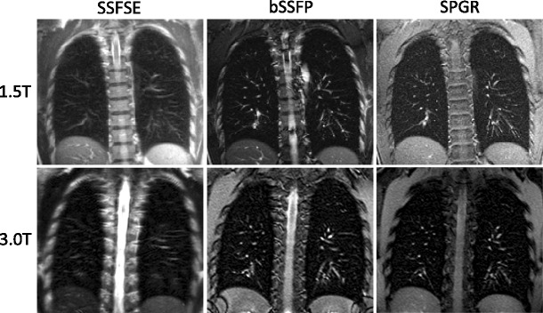
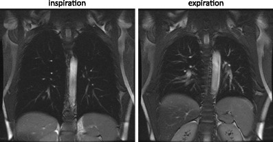
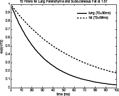
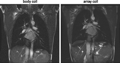
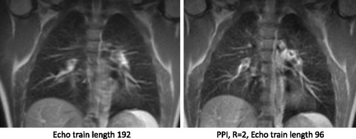
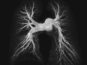
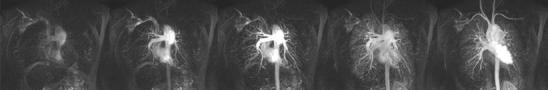
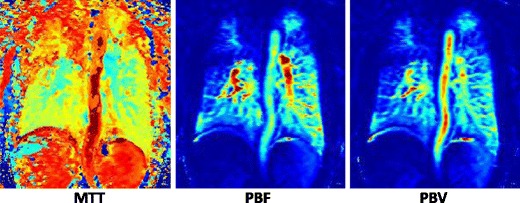
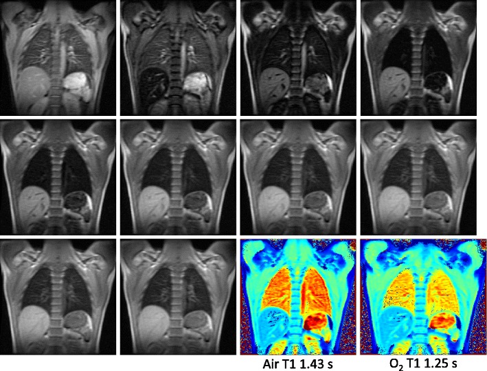
Proton magnetic resonance imaging (MRI) has recently emerged as a clinical tool to image the lungs. This paper outlines the current technical aspects of MRI pulse sequences, radiofrequency (RF) coils and MRI system requirements needed for imaging the pulmonary parenchyma and vasculature. Lung MRI techniques are presented as a "technical toolkit", from which MR protocols will be composed in the subsequent papers for comprehensive imaging of lung disease and function (parts 2 and 3). This paper is pitched at MR scientists, technicians and radiologists who are interested in understanding and establishing lung MRI methods. Images from a 1.5 T scanner are used for illustration of the sequences and methods that are highlighted. Main Messages • Outline of the hardware and pulse sequence requirements for proton lung MRI • Overview of pulse sequences for lung parenchyma, vascular and functional imaging with protons • Demonstration of the pulse-sequence building blocks for clinical lung MRI protocols.


Figures

Fig. 1
Fig. 1
1.5 T vs 3 T anatomical image SSFSE, bSSFP and SPGR
Fig. 2
Fig. 2
Balanced steady state free precession (bSSFP) images at full inspiration and full expiration
Fig. 3
Fig. 3
T2 filter for SSFSE
Fig. 4
Fig. 4
Body coil vs array coil
Fig. 5
Fig. 5
SSFSE with and without parallel imaging
Fig. 6
Fig. 6
UTE at 1.5 T in a patient with interstitial lung disease
Fig. 7
Fig. 7
Breath-hold CE pulmonary MRA
Fig. 8
Fig. 8
Time-resolved bolus passage TRICKS data set
Fig. 9
Fig. 9
Parametric maps of perfusion
Fig. 10
Fig. 10
A series of images acquired during inversion recovery at different inversion times with a Look-Locker sequence and the resulting T1 maps obtained breathing air and oxygen

References

    1. Biederer J, Beer M, Hirsch W, Wild J, Fabel M, Puderbach M et al (2012) MRI of the lung (2/3). Why… when… how? Insights Imaging, epub Feb 13; doi:10.1007/s13244-011-0146-8 - PMC - PubMed
    1. Biederer J, Mirsadraee S, Beer M, Molinari F, Hintze C, Bauman G et al (2012) MRI of the lung (3/3)—current applications and future perspectives. Insights Imaging. doi:10.1007/s13244-011-0142-z - PMC - PubMed
    1. Muller CJ, Loffler R, Deimling M, Peller M, Reiser M. J Magn Reson Imaging. 2001;14(2):164–168. doi: 10.1002/jmri.1167. - DOI - PubMed
    1. Anjorin A, Schmidt H, Posselt HG, Smaczny C, Ackermann H, Deimling M, Vogl TJ, Abolmaali N. Eur Radiol. 2008;18(6):1153–1161. doi: 10.1007/s00330-008-0884-2. - DOI - PubMed
    1. Arnold JF, Morchel P, Glaser E, Pracht ED, Jakob PM. Magn Reson Med. 2007;58(6):1092–1098. doi: 10.1002/mrm.21424. - DOI - PubMed