

Density is in the eye of the beholder: visual versus semi-automated assessment of breast density on standard mammograms
- PMID: 22696002
- PMCID: PMC3292640
- DOI: 10.1007/s13244-011-0139-7
Density is in the eye of the beholder: visual versus semi-automated assessment of breast density on standard mammograms
Abstract
Objectives: Visual inspection is generally used to assess breast density. Our study aim was to compare visual assessment of breast density of experienced and inexperienced readers with semi-automated analysis of breast density.
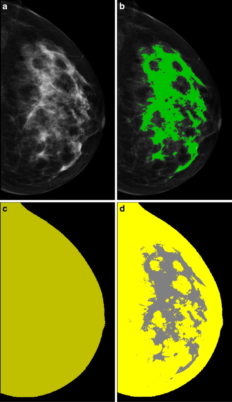
Methods: Breast density was assessed by an experienced and an inexperienced reader in 200 mammograms and scored according to the quantitative BI-RADS classification. Breast density was also assessed by dedicated software using a semi-automated thresholding technique. Agreement between breast density classification of both readers as well as agreement between their assessment versus the semi-automated analysis as reference standard was expressed as the weighted kappa value.
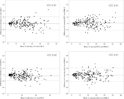
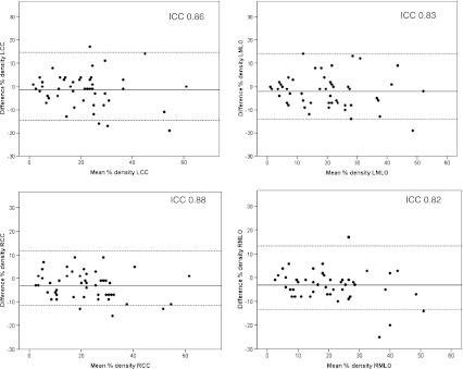
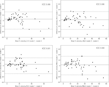
Results: Using the semi-automated analysis, agreement between breast density measurements of both breasts in both projections was excellent (ICC >0.9, P < 0.0001). Reproducibility of the semi-automated analysis was excellent (ICC >0.8, P < 0.0001). The experienced reader correctly classified the BI-RADS breast density classification in 58.5% of the cases. Classification was overestimated in 35.5% of the cases and underestimated in 6.0% of the cases. Results of the inexperienced reader were less accurate. Agreement between the classification of both readers versus the semi-automated analysis was considered only moderate with weighted kappa values of 0.367 (experienced reader) and 0.232 (inexperienced reader).
Conclusion: Visual assessment of breast density on mammograms is inaccurate and observer-dependent.
Figures
References
-
- Wolfe JN. Breast patterns as an index of risk for developing breast cancer. Am J Roentgenol. 1976;126:1130–1139. - PubMed
-
- Carney PA, Miglioretti DL, Yankaskas BC, Kerlikowske K, Rosenberg R, et al. Individual and combined effects of age, breast density, and hormone replacement therapy use on the accuracy of screening mammography. Ann Intern Med. 2003;138:168–175. - PubMed
LinkOut - more resources
Full Text Sources
