. 2012 Apr;3(2):181-7.
doi: 10.1007/s13244-011-0145-9. Epub 2012 Feb 4.

Multidetector CT cystography for imaging colovesical fistulas and iatrogenic bladder leaks

Affiliations

Multidetector CT cystography for imaging colovesical fistulas and iatrogenic bladder leaks

Massimo Tonolini et al. Insights Imaging. 2012 Apr.

Abstract

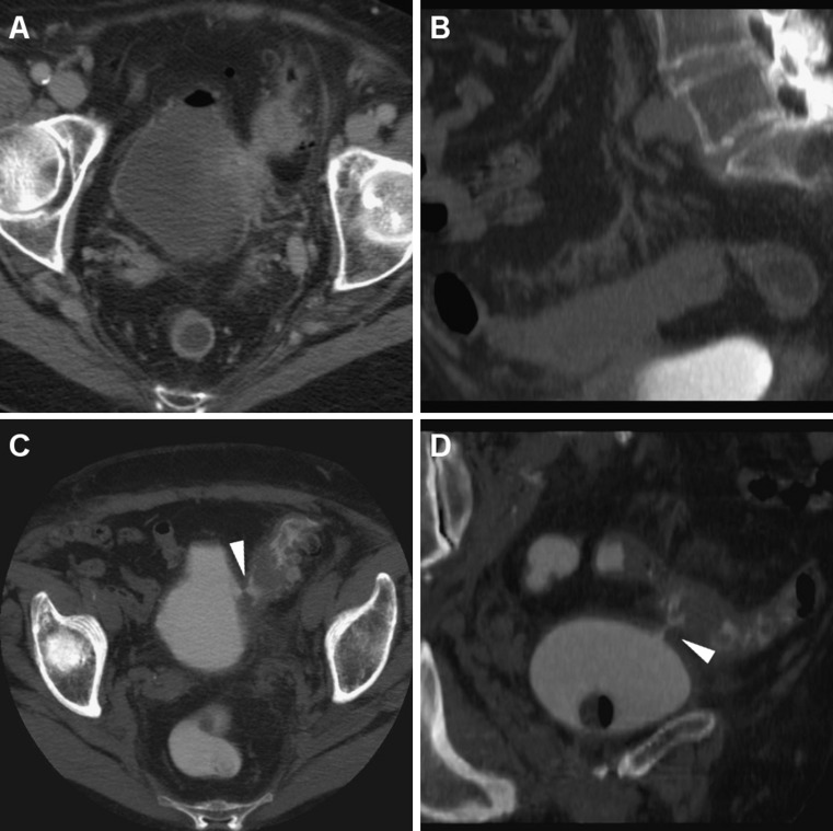
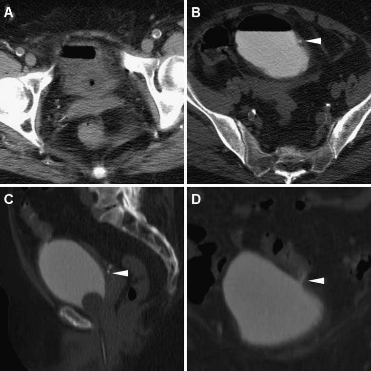
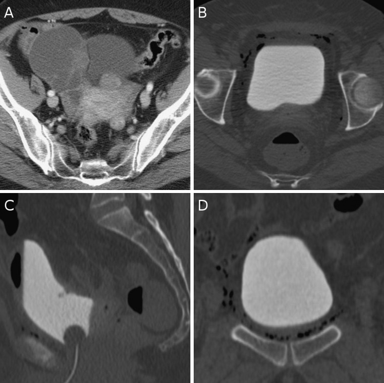
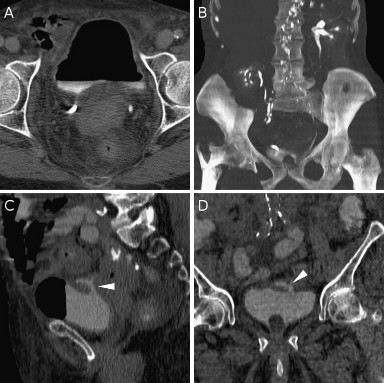
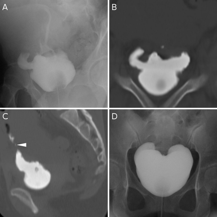
Multidetector computed tomography (MDCT) cystography currently represents the modality of choice to image the urinary bladder in traumatized patients. In this review we present our experience with MDCT cystography applications outside the trauma setting, particularly for diagnosing bladder fistulas and leaks. A detailed explanation is provided concerning exam preparation, acquisition technique, image reconstruction and interpretation. Colovesical fistulas most commonly occur as a complication of sigmoid diverticular disease, and often remain occult after extensive diagnostic work-up including cystoscopy and contrast-enhanced CT. We consistently achieved accurate preoperative visualization of colovesical fistulas using MDCT cystography. Urinary leaks and injuries represent a non-negligible occurrence after pelvic surgery, particularly obstetric and gynaecological procedures: in our experience MDCT cystography is useful to investigate iatrogenic bladder leaks or fistulas. In our opinion, MDCT cystography should be recommended as the first line modality for direct visualization or otherwise confident exclusion of both spontaneous enterovesical fistulas and bladder injuries following instrumentation procedures, obstetric or surgical interventions. Main Messages • Explanation of exam preparation, acquisition technique, image reconstruction and interpretation. • Preoperative visualization of colovesical fistulas, usually secondary to sigmoid diverticulitis. • Visualization or exclusion of iatrogenic bladder injuries following instrumentation or surgery.


Figures

Fig. 1
Fig. 1
A 79-year-old woman complaining of hypogastric pain, dysuria and fecaluria. Cystoscopy did not identify fistulous orifices. Abdomino-pelvic CT showed sigmoid diverticulosis with mild signs of perivisceral and fascial inflammation, focally adherent to the thickened left bladder wall (a). Some air in the bladder lumen without previous catheterization suggested a probable colovesical fistula, which was not directly visualized during excretory phase acquisition (focused sagittal MIP image in b). At MDCT cystography, significant leakage of diluted contrast agent in the sigmoid colon was observed through a short fistula, effectively depicted (arrowheads) on axial (c) and oblique-coronal reformatted (d) images. Elective surgery included segmental resection of the sigmoid colon, resection and repair of the bladder dome
Fig. 2
Fig. 2
An 80-year-old woman undergoing routine body CT for long-standing, unexplained fever. Air was present in the urinary bladder, with Foley catheter balloon in place (a). MDCT cystography (axial, sagittal reformatted and oblique coronal images in b, c, and d respectively) obtained adequate bladder distension with instillation of 300 ml diluted contrast medium, and visualization (arrowheads) of a subtle, initially unsuspected colovesical fistula due to colonic diverticular disease
Fig. 3
Fig. 3
A 61-year-old patient undergoing laparotomic surgery for Stage II (FIGO) bilateral ovarian cystoadenocarcinoma (preoperative staging CT image in a). MDCT cystography was performed 4 h after surgery because the gynaecological surgeon suspected intraoperative bladder rupture. Multiplanar assessment (axial, sagittal and coronal images in a, b and c, respectively) with adequate bladder filling detected some extraperitoneal air without extravasated contrast in the perivesical spaces, allowed confident exclusion of bladder rupture with urine leak and possible vesico-vaginal fistula. The patient recovered well and was discharged from hospital
Fig. 4
Fig. 4
An elderly, 86-year-old woman with multiple medical problems, hospitalized for right femur fracture. CT urography requested for clinical suspicion of acute pyelonephritis detected extensive presence of air in the bladder (a), but no extravesical urine leak was identified even on coronal MIP reformations (b). MDCT cystography (sagittal and coronal reformatted images in c and d, respectively) clearly opacified a tear in the upper posterior aspect of the bladder (arrowheads), which was retrospectively attributed to difficult emergency catheterization manoeuvres
Fig. 5
Fig. 5
A 23-year-old female investigated with conventional cystography by the attending radiologist, shortly after troublesome Caesarean section, with confirmation of bladder rupture including a fairly large right-sided pseudo-diverticular outpouching (a). MDCT cystography was requested for clarification and operative treatment planning, and revealed another large extravasation on the left upper bladder aspect (b) and fistulization from the bladder dome towards the ileum (arrowhead in c). After surgical repair, postoperative configuration of the urinary bladder was documented with radiographic cystography (d)

References

    1. Lis LE, Cohen AJ. CT cystography in the evaluation of bladder trauma. J Comput Assist Tomogr. 1990;14:386–389. doi: 10.1097/00004728-199005000-00012. - DOI - PubMed
    1. Peng MY, Parisky YR, Cornwell EE, 3rd, et al. CT cystography versus conventional cystography in evaluation of bladder injury. AJR Am J Roentgenol. 1999;173:1269–1272. - PubMed
    1. Morgan DE, Nallamala LK, Kenney PJ, et al. CT cystography: radiographic and clinical predictors of bladder rupture. AJR Am J Roentgenol. 2000;174:89–95. - PubMed
    1. Vaccaro JP, Brody JM. CT cystography in the evaluation of major bladder trauma. Radiographics. 2000;20:1373–1381. - PubMed
    1. Deck AJ, Shaves S, Talner L, et al. Computerized tomography cystography for the diagnosis of traumatic bladder rupture. J Urol. 2000;164:43–46. doi: 10.1016/S0022-5347(05)67445-9. - DOI - PubMed

LinkOut - more resources