. 2012 Jun;3(3):229-46.
doi: 10.1007/s13244-012-0152-5. Epub 2012 Mar 27.

Paediatric multi-detector row chest CT: what you really need to know

Affiliations

Paediatric multi-detector row chest CT: what you really need to know

Carolyn Young et al. Insights Imaging. 2012 Jun.

Abstract

Background: The emergence of multi-detector row CT (MDCT) has established and extended the role of CT especially in paediatric chest imaging. This has altered the way in which data is acquired and is perceived as the 'gold standard' in the detection of certain chest pathologies. The range of available post-processing tools provide alternative ways in which CT images can be manipulated for review and interpretation in order to enhance diagnostic accuracy.

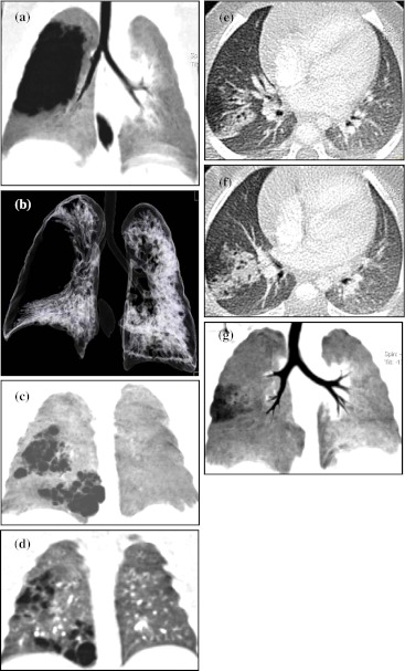
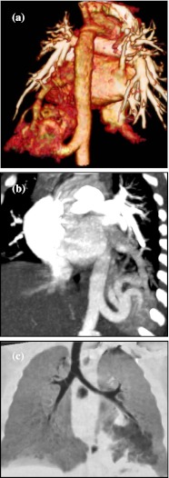
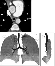
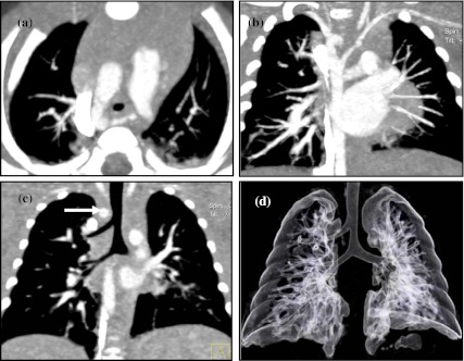
Methodology: Paediatric imaging technique/protocol together with radiation dose reduction is discussed in detail. The use of different post-processing tools to best demonstrate the wide range of important congenital anomalies and thoracic pathologies is outlined and presented pictorially.

Conclusion: MDCT with its isotropic resolution and fast imaging acquisition times reduces the need for invasive diagnostic investigations. However, users must be vigilant in their imaging techniques to minimise radiation burden, whilst maintaining good image quality. Main Messages • CT examinations should be clinically justified by the referring clinician and radiologist. • MDCT is invaluable for evaluating the central airway, mediastinal structures and lung parenchyma. • MDCT is more sensitive than plain radiographs in detection of structural changes within the lungs.


Figures

Fig. 1
Fig. 1
Altering the reconstruction algorithm from soft tissue (B30) to a bony filter (B60) increases the edge enhancement between tissue interfaces, resulting in increase in image sharpness, as demonstrated in b when compared to a, when viewed at the same parameter of W1200, C-600. Altering the viewing parameter to W400, C40 on the B30 algorithm highlights the mediastinum and great vessels (c). Scanning parameters: 100 kV, 36 eff mAs, 30 ref mAs, 1.20 CTDIvol, 20 DLP
Fig. 2
Fig. 2
The position of the ascending aorta or pulmonary artery cannot be distinguished on this axial pre-monitoring image (a) in a 1-month-old child with a large thymus and lack of mediastinal body fat, making it difficult to accurately place the ROI for automatic scan triggering. Post contrast enhancement positions of the great vessels are clearly seen in b. Scanning parameters: 80 kV, 10 mAs
Fig. 3
Fig. 3
a, b Different rendering software highlights different anatomical structures, as illustrated in these images of a 2-month-old child with congenital emphysema. a Using the MaxIP application, the parenchymal vasculature is clearly seen in the coronal and axial images (i and ii), even within the affected lobes. b Coronal (iii) and axial (iv) MinIP reformatted images show the central airway together with segmental air trapping, easily visualised within both lower lobes. Scanning parameters: 80 kV, 83 eff mAs, 60 ref mAs, 1.23 CTDIvol, 18 DLP
Fig. 4
Fig. 4
a VRT images (iiii) demonstrate the presence of a vascular ring, and its relationship with the trachea is seen in the air bronchogram (iv). The trachea is significantly compressed by the dominant right aortic arch and the aberrant left subclavian artery passing behind the trachea and oesophagus. Scanning parameters: 80 kV, 35 eff mAs, 112 ref mAs, 0.44 CTDIvol, 6 DLP. b A surface rendered air bronchogram of the tracheobronchial tree in a 5-year-old child with bronchial atresia in the affected left upper lobe. Scanning parameters: 100 kV, 46 eff mAs, 50 ref mAs, 1.53 CTDIvol, 34 DLP
Fig. 5
Fig. 5
Surface shaded display of the intra-luminal surface of the trachea (ab) seen superiorly towards the carina. The images show marked AP compression of the trachea. Axial image (c) confirms the tracheal compression between the innominate artery anteriorly and a dilated oesophagus posteriorly. Scanning parameters: 80 kV, 35 eff mAs, 150 ref mAs, 0.58 CTDIvol, 9 DLP
Fig. 6
Fig. 6
The MaxIP image (a) in a 1-month-old child shows lobar over-inflation with herniation of the lung parenchyma across the midline and also stretching of the left main bronchus. The MinIP image (b) shows generalised reduced pruning and attenuation of lung vessels in the affected lobe. Scanning parameters: 80 kV, 44 eff mAs, 60 ref mAs, 0.65 CTDIvol, 11 DLP
Fig. 7
Fig. 7
Type 1 CPAM consists of a large cyst with multiple smaller cysts seen on the coronal MinIP image (a) causing mass effect with distortion to the remainder of the right lung parenchyma with mediastinal shift seen on the air bronchogram (b). Scanning parameters: 80 kV, 60 eff mAs, 60 ref mAs, 0.87 CTDIvol, 16 DLP. Type 2 CPAM with multiple cystic lesions in the right lower lobe is better defined on the coronal MinIP image (c) than on the MPR image (d). Scanning parameters: 80 kV, 51 eff mAs, 60 ref mAs, 0.76 CTDIvol, 12 DLP. Type 3 CPAM axial images (e, f) show an area of consolidation with adjacent region of hyperinflation. There is tubular lucency seen within this apical segment of the right lower lobe with air-trapping within the lesion as shown on the MinIP image (g). Scanning parameters: 80 kV, 60 eff mAs, 60 ref mAs, 0.90 CTDIvol, 18 DLP
Fig. 8
Fig. 8
Posterior VRT image (a) of an 11-month-old child demonstrates an extra-lobar sequestration with a systemic arterial supply arising from the abdominal aorta and venous drainage into the hepatic veins. Differentiation between normal lung parenchyma and the sequestrated lobe is seen on both the coronal air bronchogram image (b) and MaxIP image (c). Scanning parameters: 80 kV, 54 eff mAs, 60 ref mAs, 0.80 CTDIvol, 18 DLP
Fig. 9
Fig. 9
Both the posterior-oblique VRT (a) and the coronal MaxIP (b) images demonstrate an intra-lobar sequestration in the left lower lobe of a 3-month-old child. There is a large (systemic) arterial feeding vessel originating from the abdominal aorta. Venous drainage of the sequestrated lobe is conventional into the pulmonary vein. Abnormal tissue can be seen within the left lower lobe on the MinIP image (c). Scanning parameters: 80 kV, 47 eff mAs, 60 ref mAs, 0.70 CTDIvol, 13 DLP
Fig. 10
Fig. 10
MaxIP images (a, b) demonstrate an anomalous curvilinear right lower pulmonary vein draining into the inferior vena cava just above the hemi-diaphragm, a classic sign of Scimitar syndrome, in a 10-year-old child. Previous metallic embolisation coils are seen on both images. Scanning parameters: 100 kV, 64 eff mAs, 150 ref mAs, 7.15 CTDIvol, 128 DLP
Fig. 11
Fig. 11
A 12-year-old child with congenital tracheal stenosis associated with complete cartilaginous ring and ‘O’-shaped trachea (in the axial plane) (a). The trachea is long with an obtuse carina angle that ends at the T6 level (MinIP image, b), whilst the stenotic tracheal length is best depicted on the sagittal MinIP image (c). Scanning parameters: 100 kV, 44 eff mAs, 180 ref mAs, 5.70 CTDIvol, 135 DLP
Fig. 12
Fig. 12
Incidental finding of an accessory right apical bronchus (pig bronchus) in a 3-month-old child with associated right aortic arch and an aberrant left subclavian artery (MinIP images, a and b), forming a vascular ring around the trachea, with tracheal compression (c, arrow). The pig bronchus is clearly demonstrated on coronal MPR image (c) and on the CT bronchogram image (d). Note also the widened carina angle. Scanning parameters: 80 kV, 86 eff mAs, 86 ref mAs, 2.72 CTDIvol, 38 DLP
Fig. 13
Fig. 13
MaxIP image (a) of a 5-year-old child shows left upper lobe over-inflation with classical pruning of attenuated pulmonary vasculature. A mucoid plug can be seen (arrow) with a proximal bronchcoele in keeping with bronchial atresia (b, c). Scanning parameters: 100 kV, 46 eff mAs, 50 ref mAs, 1.53 CTDIvol, 34 DLP
Fig. 14
Fig. 14
Coronal and axial images show a well-defined water density lesion lying within the mediastinum. There is no systemic arterial supply to the lesion, and it is not connected to the tracheobronchial tree. Appearances are compatible with a mediastinal bronchogenic cyst. Scanning parameters: 80 kV, 57 eff mAs, 60 ref mAs, 0.85 CTDIvol, 13 DLP
Fig. 15
Fig. 15
Three types of bronchial dilatation can be seen in patients with bronchiectasis, and their appearance on CT can be classified into a cylindrical, b varicose and c saccular shapes
Fig. 16
Fig. 16
Axial MaxIP images in a 12-year-old child with advanced cystic fibrosis show ‘tree-in-a-bud’ appearances related to centrilobular exudative bronchiolectasis. Scanning parameters: 100 kV, 79 eff mAs, 42 ref mAs, 2.63 CTDIvol, 70 DLP
Fig. 17
Fig. 17
HRCT shows ground-glass opacification with interlobular septal fluid thickening giving the ‘crazy paving’ appearance is illustrated in a 1.5-year-old immuno-compromised child with cytomegalovirus (CMV) infection. Scanning parameters: 100 kV, 41 eff mAs, 30 ref mAs, 1.38 CTDIvol, 25 DLP
Fig. 18
Fig. 18
VRT (a) and MaxIP images (b) in a 1.5-year-old child show a (right-sided dominant) double aortic arch that encircles the trachea and oesophagus causing narrowing of the trachea at this level and extends down to the carina seen on the MinIP image (c). Scanning parameters: 80 kV, 39 eff mAs, 150 ref mAs, 0.67 CTDIvol, 9 DLP
Fig. 19
Fig. 19
a Posterior oblique VRT image of a 2-month-old child shows a right-sided aortic arch with mirror branching pattern demonstrated. Scanning parameters: 80 kV, 62 mAs, 0.70 CTDIvol, 8 DLP. b Axial MaxIP image (i) of a 2-year-old child shows a right dominant aortic arch with a Kommerell’s diverticulum and an aberrant left subclavian artery seen on the VRT image (ii). A patent arterial duct connects to the left pulmonary artery, completing a vascular ring around the trachea that is mildly compressed due to a dilated oesophagus seen in (i). Scanning parameters: 80 kV, 33 eff mAs, 150 ref mAs, 1.15 CTDIvol, 20 DLP
Fig. 20
Fig. 20
CT images (a, b) show severe tracheobronchial compression in a baby with biphasic stridor. An aberrant left pulmonary artery is seen arising from the right pulmonary artery (a), creating a vascular sling around the trachea as it passes posteriorly causing almost complete occlusion of the distal trachea (b), with marked narrowing of the left main bronchus. Scanning parameters: 80 kV, 58 eff mAs, 60 ref mAs, 0.86 CTDIvol, 14 DLP
Fig. 21
Fig. 21
MDCT images of a child with a single pulmonary vein with severe narrowing at the junction where it enters the posterior aspect of the left atrium. This can be seen clearly on both the axial MaxIP image (a) and on the VRT image (b). MDCT is the investigation of choice in patients with venous stenosis due to the superior spatial resolution of CT over MRI. Scanning parameters: 80 kV, 38 eff mAs, 130 ref mAs, 0.66 CTDIvol, 8 DLP

References

    1. Sundaram B, Chughtai AR, Kazerooni EA. Multi-detector high resolution computed tomography of the lungs. J Thorac Imaging. 2010;25:125–141. doi: 10.1097/RTI.0b013e3181d9ca37. - DOI - PubMed
    1. Toma P, Rizzo F, Stagnaro N, Magnano G, Granata C. Multislice CT in congenital bronchopulmonary malformations in children. Radiol Med. 2010;116:133–151. doi: 10.1007/s11547-010-0582-4. - DOI - PubMed
    1. Lee EY, Boiselle PM, Shamberger RC. Multidetector CT and 3D imaging: preoperative evaluation of thoracic vascular and tracheobronchial anomalies and abnormalities in pediatric patients. J Pediatr Surg. 2010;45:811–821. doi: 10.1016/j.jpedsurg.2009.12.013. - DOI - PubMed
    1. Aziz ZA, Padley SP, Hansell DM. CT techniques for imaging the lung: recommendations for multislice and single slice computed tomography. Eur J Radiol. 2004;52:119–136. doi: 10.1016/j.ejrad.2004.01.005. - DOI - PubMed
    1. Boiselle PM, Lee KS, Ernst A. Multidetector CT of the central airway. J Thorac Imaging. 2005;20:186–195. doi: 10.1097/01.rti.0000171624.84951.f2. - DOI - PubMed

LinkOut - more resources