. 2012 Jun;3(3):247-50.
doi: 10.1007/s13244-012-0168-x. Epub 2012 May 1.

Hypertrophic pyloric stenosis: tips and tricks for ultrasound diagnosis

Affiliations

Hypertrophic pyloric stenosis: tips and tricks for ultrasound diagnosis

Sílvia Costa Dias et al. Insights Imaging. 2012 Jun.

Abstract

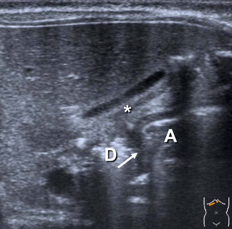
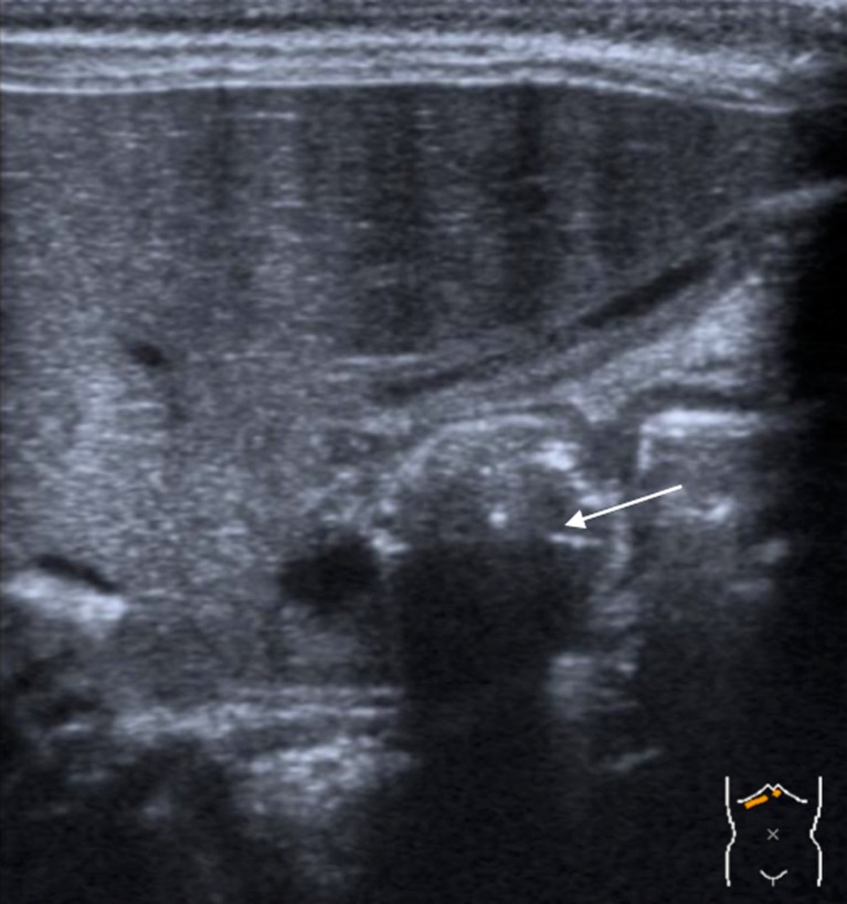
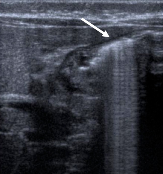
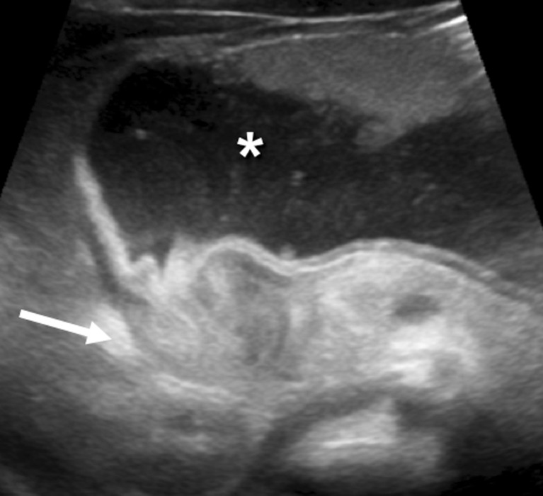
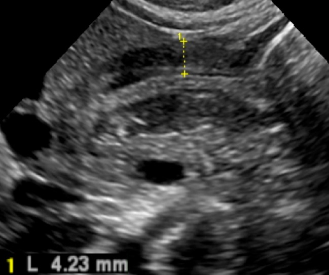
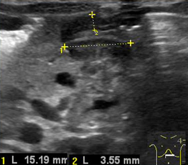
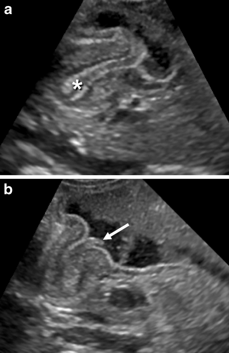
We describe a systematic approach to the ultrasound (US) examination of the antropyloric region in children. US is the modality of choice for the diagnosis of hypertrophic pyloric stenosis (HPS). The imaging features of the normal pylorus and the diagnostic findings in HPS are reviewed and illustrated in this pictorial essay. Common difficulties in performing the examination and tips to help overcome them will also be discussed. Main Messages • Hypertrophic Pyloric Stenosis is defined by thickening of the muscular layer and failure in relaxation of the pyloric canal. • The main diagnostic criterion is a measurement of more than 3mm in thickness of the muscular layer. • Abnormal elongation of the canal is characterised as greater than 12 mm in length.


Figures

Fig. 1
Fig. 1
The pylorus (arrow) between the gastric antrum (A) and the duodenum (D) lying posterior to the gallbladder (*)
Fig. 2
Fig. 2
Passage of the gastric content through the pylorus, distending the antropyloric region (arrow)
Fig. 3
Fig. 3
The stomach distended with gas (arrow)
Fig. 4
Fig. 4
The distended stomach (*), posteriorly displacing the pylorus (arrow), which resembles the appearance of the uterine cervix
Fig. 5
Fig. 5
The hypertrophied muscular layer
Fig. 6
Fig. 6
Abnormal elongation of the pyloric canal (measure 1)
Fig. 7
Fig. 7
a The double layer of thickened mucosa (*), b protruding through the antrum

References

    1. Ohshiro K, Puri P. Pathogenesis of infantile hypertrophic pyloric stenosis: recent progress. Pediatr Surg Int. 1998;13:43–252. - PubMed
    1. Hiorns MP. Gastrointestinal tract imaging in children: current techniques. Pediatr Radiol. 2011;41:42–54. doi: 10.1007/s00247-010-1743-2. - DOI - PubMed
    1. Panteli C. New insights into the pathogenesis of infantile pyloric stenosis. Pediatr Surg Int. 2009;25:1043–1052. doi: 10.1007/s00383-009-2484-x. - DOI - PubMed
    1. Chandran L, Chitkara M. Vomiting in children: reassurance, red flag, or referral? Pediatr Rev. 2008;29:183–192. doi: 10.1542/pir.29-6-183. - DOI - PubMed
    1. Hernanz-Schulman M. Infantile hypertrophic pyloric stenosis. Radiology. 2003;227:319–331. doi: 10.1148/radiol.2272011329. - DOI - PubMed

LinkOut - more resources