. 2012 Jun;3(3):251-63.
doi: 10.1007/s13244-012-0154-3. Epub 2012 Mar 17.

Small bowel MR enterography: problem solving in Crohn's disease

Affiliations

Small bowel MR enterography: problem solving in Crohn's disease

Nyree Griffin et al. Insights Imaging. 2012 Jun.

Abstract

Magnetic resonance enterography (MRE) is fast becoming the first-line radiological investigation to evaluate the small bowel in patients with Crohn's disease. It can demonstrate both mural and extramural complications. The lack of ionizing radiation, together with high-contrast resolution, multiplanar capability and cine-imaging make it an attractive imaging modality in such patients who need prolonged follow-up. A key question in the management of such patients is the assessment of disease activity. Clinical indices, endoscopic and histological findings have traditionally been used as surrogate markers but all have limitations. MRE can help address this question. The purpose of this pictorial review is to (1) detail the MRE protocol used at our institution; (2) describe the rationale for the MR sequences used and their limitations; (3) compare MRE with other small bowel imaging techniques; (4) discuss how MRE can help distinguish between inflammatory, stricturing and penetrating disease, and thus facilitate management of this difficult condition. Main Messages • MR enterography (MRE) is the preferred imaging investigation to assess Crohn's disease. T2-weighted, post-contrast and diffusion-weighted imaging (DWI) can be used. • MRE offers no radiation exposure, high-contrast resolution, multiplanar ability and cine imaging. • MRE can help define disease activity, a key question in the management of Crohn's disease. • MRE can help distinguish between inflammatory, stricturing and penetrating disease. • MRE can demonstrate both mural and extramural complications.


Figures
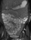
Fig. 1
Fig. 1
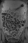
Coronal True FISP image: normal bowel. The ‘black boundary’ artefact may be confused with bowel wall thickening (arrows)
Fig. 2
Fig. 2
Coronal T1 fat-saturated post-contrast image: normal bowel wall shows mild homogeneous enhancement
Fig. 3
Fig. 3
Coronal fat-saturated HASTE image: normal bowel. Intraluminal flow voids (arrow) are seen, as this sequence is sensitive to fluid motion
Fig. 4
Fig. 4
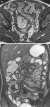
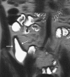
Example of mural thickening in active Crohn’s disease: a axial True FISP image shows mural thickening in the distal ileum (arrow); b coronal True FISP in a different patient (15 years old) shows extensive jejunal small bowel wall thickening (arrows); this distribution of disease is less common than distal/terminal ileum. Note the fibrofatty proliferation in the adjacent mesentery
Fig. 5
Fig. 5
Example of pseudosacculation: coronal True FISP image shows two long skip lesions in the mid to distal ileum (asterisk) demonstrating mural thickening, luminal narrowing and prominence of the vasa recta (arrowheads) with small nodes seen within the mesentery. There are multiple pseudosacculations (arrows) on the antimesenteric border due to asymmetric involvement of the bowel wall. Mesenteric fibrofatty proliferation separates the involved bowel loops. Appearances suggest a mixture of active and fibrostenotic disease
Fig. 6
Fig. 6
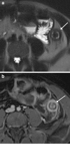
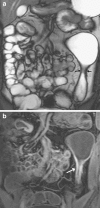
Patient with active distal ileal Crohn’s disease: a) axial HASTE image shows bowel wall is of intermediate signal (arrow) due to submucosal oedema. Mesenteric oedema (asterisk) is also noted; b) corresponding axial T1 fat-saturated post-contrast image shows avid stratified pattern of mural enhancement (arrow)
Fig. 7
Fig. 7
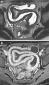
Patient with active distal colitis: a axial HASTE image shows very high signal in the submucosa in keeping with oedema (arrow); b corresponding axial T1 fat-saturated post-contrast image shows avid stratified pattern of mural enhancement (arrow)
Fig. 8
Fig. 8
Example of fibrostenotic disease: coronal HASTE image shows a stricture in the neoterminal ileum in a patient with previous ileocolic resection; this is of intermediate to low signal due to little mural oedema (arrows); there is pre-stenotic dilatation
Fig. 9
Fig. 9
Patient with fibrostenotic disease: a coronal True FISP image showing short fibrotic stricture (arrows) in the mid descending colon with marked pre-stenotic dilatation; b corresponding homogeneous mural enhancement is seen in this skip lesion (arrow)
Fig. 10
Fig. 10
Example of comb sign in active inflammation: coronal True FISP image shows multiple linear low signal structures extending to the bowel wall in keeping with engorged vasa recta (arrows) supplying the thickened terminal ileum (asterisk). Note the fibrofatty proliferation within the adjacent mesentery, separating the inflamed terminal ileum from adjacent loops of bowel, with small mesenteric nodes also present
Fig. 11
Fig. 11
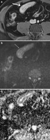
Example of DWI in active inflammation: a axial T1 fat-saturated post-contrast image shows thickened enhancing ileum with typical stratified enhancement pattern (arrow); b corresponding DWI (b = 800) and c ADC map confirm restricted diffusion with persistent high signal (arrow) on the DWI and low signal (arrow) on the ADC map (ROI = 1,200)
Fig. 12
Fig. 12
Complications of penetrating disease: a coronal True FISP image showing an enteroenteric fistula (arrows) between an inflamed segment of mid ileum (white asterisk) and non-inflamed terminal ileum (black asterisk); b Axial True FISP image in a different patient showing an enterocutaneous fistula (arrows) between a loop of inflamed thickened small bowel (asterisk) and skin; c axial True FISP image in a different patient showing the ‘star’ sign between adjacent loops of bowel, highly suggestive of enteroenteric fistulae (arrows); d coronal T1 fat saturated post contrast image in a different patient again showing multiple converging enhancing loops of small bowel suggestive of enteroenteric fistulae (arrows)
Fig. 13
Fig. 13
Complications of penetrating disease: coronal T1 fat-saturated post-contrast image showing multiple enhancing skip lesions (arrows) and enhancing mesenteric phlegmon (asterisk)
Fig. 14
Fig. 14
Complication of penetrating disease: patient with active colitis showing circumferential mural thickening and stratified mural enhancement of the proximal colon (asterisk), complicated by an ileopsoas abscess (arrows)
Fig. 15
Fig. 15
Complication of penetrating disease: patient with local perforation due to penetrating Crohn’s disease of the proximal colon—a thick walled air-containing cavity is seen extending into the anterior abdominal wall (arrows)
Fig. 16
Fig. 16
Typical example of response to treatment with a biologic: a axial T1 fat-saturated post-contrast image shows a thickened avidly enhancing segment of midileum with stratified pattern of enhancement (arrows) in keeping with active inflammation; b axial T1 fat-saturated post-contrast image following 8 months of infliximab shows inflammation has been downstaged with reduction in mural thickening and enhancement which is now homogeneous (arrows)

Similar articles

Cited by

References

    1. Loftus EV., Jr Clinical epidemiology of inflammatory bowel disease: incidence, prevalence, and environmental influences. Gastroenterology. 2004;126:1504–1517. doi: 10.1053/j.gastro.2004.01.063. - DOI - PubMed
    1. Assche G, Dignass A, Panes J, Beaugerie L, Karagiannis J, Allez M, Ochsenkühn T, Orchard T, Rogler G, Louis E, Kupcinskas L, Mantzaris G, Travis S, Stange E, European Crohn’s and Colitis Organisation (ECCO) The second European evidence-based Consensus on the diagnosis and management of Crohn’s disease: Definitions and diagnosis. J Crohns Colitis. 2010;4:7–27. doi: 10.1016/j.crohns.2009.12.003. - DOI - PubMed
    1. Hafeez R, Punwani S, Boulos P, Bloom S, McCartney S, Halligan S, Taylor SA. Diagnostic and therapeutic impact of MR enterography in Crohn’s disease. Clin Radiol. 2011;66:1148–1158. doi: 10.1016/j.crad.2010.12.018. - DOI - PubMed
    1. Cronin CG, Lohan DG, Mhuircheartaigh JN, McKenna D, Alhajeri N, Roche C, Murphy JM. MRI small-bowel follow-through: prone versus supine patient positioning for best small-bowel distention and lesion detection. AJR Am J Roentgenol. 2008;191:502–506. doi: 10.2214/AJR.07.2338. - DOI - PubMed
    1. Lemberg DA, Issenman RM, Cawdron R, Green T, Mernagh J, Skehan SJ, et al. Positron emission tomography in the investigation of pediatric inflammatory bowel disease. Inflamm Bowel Dis. 2005;11:733–738. doi: 10.1097/01.MIB.0000172810.49619.cb. - DOI - PubMed

LinkOut - more resources