Fetal MR in the evaluation of pulmonary and digestive system pathology
- PMID: 22696089
- PMCID: PMC3369121
- DOI: 10.1007/s13244-012-0155-2
Fetal MR in the evaluation of pulmonary and digestive system pathology
Abstract
Background: Prenatal awareness of an anomaly ensures better management of the pregnant patient, enables medical teams and parents to prepare for the delivery, and is very useful for making decisions about postnatal treatment. Congenital malformations of the thorax, abdomen, and gastrointestinal tract are common. As various organs can be affected, accurate location and morphological characterization are important for accurate diagnosis.
Methods: Magnetic resonance imaging (MRI) enables excellent discrimination among tissues, making it a useful adjunct to ultrasonography (US) in the study of fetal morphology and pathology.
Results: MRI is most useful when US has detected or suspected anomalies, and more anomalies are detected when MRI and US findings are assessed together.
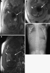
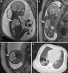
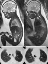
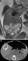
Conclusion: We describe the normal appearance of fetal thoracic, abdominal, and gastrointestinal structures on MRI, and we discuss the most common anomalies involving these structures and the role of MRI in their study.
Teaching points: • To learn about the normal anatomy of the fetal chest, abdomen, and GI tract on MRI. • To recognize the MR appearance of congenital anomalies of the lungs and the digestive system. • To understand the value of MRI when compared to US in assessing fetal anomalies.
Figures








Similar articles
-
Magnetic resonance imaging in the prenatal diagnosis of neural tube defects.Insights Imaging. 2013 Apr;4(2):225-37. doi: 10.1007/s13244-013-0223-2. Epub 2013 Mar 1. Insights Imaging. 2013. PMID: 23456749 Free PMC article.
-
Fetal Neurosonogaphy: Ultrasound and Magnetic Resonance Imaging in Competition.Ultraschall Med. 2016 Dec;37(6):555-557. doi: 10.1055/s-0042-117142. Epub 2016 Dec 15. Ultraschall Med. 2016. PMID: 27978593 English.
-
Prenatal diagnosis of congenital head, face, and neck malformations-Is complementary fetal MRI of value?Prenat Diagn. 2020 Jan;40(1):142-150. doi: 10.1002/pd.5593. Epub 2019 Nov 11. Prenat Diagn. 2020. PMID: 31664716
-
Evaluation of prenatally diagnosed structural congenital anomalies.J Obstet Gynaecol Can. 2009 Sep;31(9):875-881. doi: 10.1016/S1701-2163(16)34307-9. J Obstet Gynaecol Can. 2009. PMID: 19941713 Review. English, French.
-
Role of magnetic resonance imaging in fetuses with mild or moderate ventriculomegaly in the era of fetal neurosonography: systematic review and meta-analysis.Ultrasound Obstet Gynecol. 2019 Aug;54(2):164-171. doi: 10.1002/uog.20197. Epub 2019 Jul 11. Ultrasound Obstet Gynecol. 2019. PMID: 30549340
Cited by
-
Fetal thoracic teratomas: mediastinal or pericardial?Pediatr Radiol. 2022 Nov;52(12):2319-2328. doi: 10.1007/s00247-022-05367-x. Epub 2022 Apr 26. Pediatr Radiol. 2022. PMID: 35471666
-
Fetal Magnetic Resonance Imaging of Malformations Associated with Heterotaxy.Cureus. 2015 May 21;7(5):e269. doi: 10.7759/cureus.269. eCollection 2015 May. Cureus. 2015. PMID: 26180693 Free PMC article. Review.
-
Magnetic Resonance Imaging Diagnosis of Volvulus through Mesenteric Defect in Neonate.AJP Rep. 2016 Apr;6(2):e239-42. doi: 10.1055/s-0036-1584548. AJP Rep. 2016. PMID: 27551577 Free PMC article.
-
[Fetal magnetic resonance imaging of thoracic and abdominal malformations].Radiologe. 2013 Feb;53(2):123-9. doi: 10.1007/s00117-012-2400-6. Radiologe. 2013. PMID: 23354851 German.
References
-
- Martin C, Darnell A, Escofet C, Mellado F, Corona M. Fetal magnetic resonance imaging. Ultrasound Rev Obstet Gynecol. 2004;4:214–227.
LinkOut - more resources
Full Text Sources
Research Materials