. 2012 Sep;97(9):655-68.
doi: 10.1002/bip.22065.

Gorgon and pathwalking: macromolecular modeling tools for subnanometer resolution density maps

Affiliations

Gorgon and pathwalking: macromolecular modeling tools for subnanometer resolution density maps

Matthew L Baker et al. Biopolymers. 2012 Sep.

Abstract

The complex interplay of proteins and other molecules, often in the form of large transitory assemblies, are critical to cellular function. Today, X-ray crystallography and electron cryo-microscopy (cryo-EM) are routinely used to image these macromolecular complexes, though often at limited resolutions. Despite the rapidly growing number of macromolecular structures, few tools exist for modeling and annotating structures in the range of 3-10 Å resolution. To address this need, we have developed a number of utilities specifically targeting subnanometer resolution density maps. As part of the 2010 Cryo-EM Modeling Challenge, we demonstrated two of our latest de novo modeling tools, Pathwalking and Gorgon, as well as a tool for secondary structure identification (SSEHunter) and a new rigid-body/flexible fitting tool in Gorgon. In total, we submitted 30 structural models from ten different subnanometer resolution data sets in four of the six challenge categories. Each of our utlities produced accurate structural models and annotations across the various density maps. In the end, the utilities that we present here offer users a robust toolkit for analyzing and modeling protein structure in macromolecular assemblies at non-atomic resolutions.


Figures

Figure 1
Figure 1
Subnanometer resolution density maps. A plot of the deposited macromolecular structures in the Protein Data Bank >150KDa (blue) and the EM DataBank (green) are shown sorted by resolution. Nearly 25% of all cryo-EM and ~33% of all X-ray crystallographic density maps report resolutions between 3Å and 10Å.
Figure 2
Figure 2
Density map resolution. Cryo-EM reconstructions of GroEL at 4 different resolutions are shown on the top row. The corresponding resolutions and EMDB ID numbers are shown below. A single subunit from the X-ray crystal structure of GroEL (1SS8) is shown superimposed on the cryo-EM density map in the bottom row.
Figure 3
Figure 3
SSE detection. The results for SSE detection are shown. Helices are represented as green cylinder, β-sheets as cyan planes and the density map is shown as a transparent isosurface.
Figure 4
Figure 4
De novo models. A gallery of de novo models is shown. In the first column, the density maps are shown. In the second and third column, the resulting models from Gorgon and Pathwalking are shown, respectively. For Mm-cpn, only Gorgon was used to construct the model. The two results, grouped together in the box, were constructed by two different users in Gorgon. In the final column, the known structure is shown. All structures are rainbow colored from N- (blue) to C- (red) terminus.
Figure 5
Figure 5
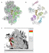
Fitting with Gorgon. Results for the fitting of eight protein subunits in T. thermophilus ribosome density map are shown in (A). In (B), the known positions of the protein subunits in the ribosome density map are shown in grey with the Gorgon fits superimposed. The Gorgon “Fit to density” interface is shown in (C).
Figure 6
Figure 6
Pathwalking versus Gorgon model building. The results for Pathwalking (green) and Gorgon (blue) model building for rotavirus VP6 are shown overlaid on the crystal structure (red). Two zoomed in regions of the models are shown overlaid on the density map to demonstrate the accuracy of model construction.

References

    1. Alberts B, Miake-Lye R. Cell. 1992;68:415–20. - PubMed
    1. Alberts B. Cell. 1998;92:291–4. - PubMed
    1. Sali A. Structure. 2003;11:1043–7. - PubMed
    1. Chiu W, Baker ML, Almo SC. Trends Cell Biol. 2006;16:144–50. - PubMed
    1. Baumeister W, Steven AC. Trends Biochem Sci. 2000;25:624–31. - PubMed

Publication types