. 2012 Aug;30(8):1624-33.
doi: 10.1002/stem.1143.

Regulation of embryonic stem cell pluripotency by heat shock protein 90

Affiliations

Regulation of embryonic stem cell pluripotency by heat shock protein 90

Eric Bradley et al. Stem Cells. 2012 Aug.

Abstract

Deciphering the molecular basis of stem cell pluripotency is fundamental to the understanding of stem cell biology, early embryonic development, and to the clinical application of regenerative medicine. We report here that the molecular chaperone heat shock protein 90 (Hsp90) is essential for mouse embryonic stem cell (ESC) pluripotency through regulating multiple pluripotency factors, including Oct4, Nanog, and signal transducer and activator of transcription 3. Inhibition of Hsp90 by either 17-N-Allylamino-17-demethoxygeldanamycin or miRNA led to ESC differentiation. Overexpression of Hsp90β partially rescued the phenotype; in particular, the levels of Oct4 and Nanog were restored. Notably, Hsp90 associated with Oct4 and Nanog in the same cellular complex and protected them from degradation by the ubiquitin proteasome pathway, suggesting that Oct4 and Nanog are potential novel Hsp90 client proteins. In addition, Hsp90 inhibition reduced the mRNA level of Oct4, but not that of Nanog, indicating that Hsp90 participates in Oct4 mRNA processing or maturation. Hsp90 inhibition also increased expression of some protein markers for mesodermal lineages, implying that Hsp90 suppresses mesodermal differentiation from ESCs. These findings support a new role for Hsp90 in maintaining ESC pluripotency by sustaining the level of multiple pluripotency factors, particularly Oct4 and Nanog.


Figures

Figure 1
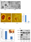
Figure 1. Hsp90 is required for stem cell pluripotency- 17-AAG induced ESC differentiation
A. Bright field images of mouse ESC colony morphology, feeder-free J1 mouse ESCs were treated with vehicle control or 250 nM 17-AAG for 48 hours. Con, vehicle treated; AAG, 250nM 17-AAG, scale bar: 30μm. B. AP staining of the mouse ESC colonies. Feeder-free mouse ESCs grown on cover slips were treated with vehicle control or 250 nM 17-AAG for 48 hours, fixed and stained with alkaline phosphatase. Left panel, typical images, scale bar: 20μm; right panel, statistic analysis of the number of AP positive colonies per cover slip. N=4, **, p<0.01. C. Teratoma formation (arrow on the inset). 17-AAG treated or untreated cells were injected intracranially into P3 mouse brains. Mice were sacrificed and relative teratoma weight was measured four weeks later, N=4, *, p<0.05. Inset shows a set of typical images of the teratoma. D. Western blot analysis of pluripotency factors. Phosphorylated Stat3 (pStat3), total Stat3 (tStat3), Nanog, and Oct4 were detected using 250nM 17-AAG- treated mouse ESC samples; β-Actin was used as loading control. Results from two different sets of samples were shown.
Figure 2
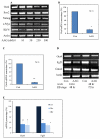
Figure 2. Hsp90 is required for stem cell pluripotency - Hsp90 miRNA led to ESC differentiation and cell death and Hsp90β prevented 17-AAG mediated pluripotency loss
A. Hsp90 miRNA reduced Oct4 and Nanog protein levels in mouse ESCs. The Hsp90α miRNA2139 and Hsp90β miRNA1201 construct, which have been proved to effectively repress Hsp90α and Hsp90β expression (Fig. S3), were electroporated into mouse ESCs. Cell lysates were assayed by Westernblot analysis for Hsp90α, Hsp90β, Oct4, and Nanog. β-actin was used as loading control. B. Hsp90 inhibition by miRNA reduced ESC differentiation. Mouse ESCs were treated as in A except that the cells were fixed for AP staining and the number of AP positive colonies was counted. Y axis represents the number of AP positive colonies per cover slip. N=4, *, p<0.05; **, p<0.01. C. Overexpression of Hsp90β prevented 17-AAG mediated ESC pluripotency loss. Feeder free mouse ESCs were infected with an Hsp90β lentiviral vector or a control (Lacz) lentiviral vector. Twenty four hours later, the culture media were supplemented with 250 nM 17-AAG and the cells were incubated for another 48 hours. Then the cells were fixed, stained, and AP positive counted. Y axis represents the number of AP positive colonies per cover slip. N=3. D. Cells were treated as in C except that cell lysates were examined by Westernblot analysis for the proteins indicated.
Figure 3
Figure 3. Hsp90 associates with Oct4 and Nanog and prevented them from degradation by the ubiquitin proteasome pathway
ESC lysates were subjected to co-IP assays. A. Cell lysate was immunoprecipated with antibodies against Hsp90β and Hsp90α, and immuno-blotted with Oct4 antibody. Beads only and pre-immune serum were used as control. B. Cell lysate was immunoprecipated with Oct4 antibody, and immuno-blotted with antibodies against Hsp90α and Hsp90β. Beads alone was used as control. C. Cell lysate was immunoprecipated with antibodies against Hsp90α and Hsp90β, and immuno-blotted with Nanog antibody. D. Cell lysate was immunoprecipated with Nanog antibody, and immuno-blotted with antibodies against Hsp90α and Hsp90β. Beads alone was used as control. E. Feeder free ESCcs were treated with 0.2μM MG132 0.5 hour prior to 250nM 17-AAG addition. Another 48 hours later, cell lysates were collected and Western blot analysis performed. Note proteasomal inhibition prevented Oct4 and Nanog protein loss mediated by 17-AAG. MG, 0.2μM MG132 treatment.
Figure 4
Figure 4
Hsp90 inhibition reduced the level of Oct4 mRNA. A. Feeder free mouse ESCs were treated for 48 hours with 17-AAG of concentrations indicated. Total RNA was collected and RT-PCR performed with primers for the mRNA of indicated genes. B. Real time quantitative PCR of Oct4 mRNA using mouse ESCs. C. Real time quantitative PCR of Oct4 mRNA on human ESCs. D. RT-PCR of Oct4 mRNA on floating EBs treated with 17-AAG for 24 hours and 48 hours. E. Densitometry quantification of the Oct4 and Fgf5 bands in D, values shown are normalized to β-actin. N=3.
Figure 5
Figure 5. Dose and time course study of the effect of Hsp90 inhibition on ESC pluripotency factors
A. Feeder free mouse ESCs were treated for 48 hours with 17-AAG of concentrations indicated. Cell lysates were collected and Western blot analysis performed for proteins indicated. Casp3, cleaved (activated) caspase 3. B. Feeder free ESCs were treated with 250 nM 17-AAG for the time period indicated. Cell lysates were subjected to Western blot analysis for proteins as in A.
Figure 6
Figure 6. Hsp90 inhibition increases markers for mesoderm differentiation
A. Mouse ESCs were treated with 250nM 17-AAG or vehicle for 48 hours, fixed or collected for immunofluorescence for markers of endoderm (AFP), ectoderm (vimentin), and mesoderm (desmin). Hoe, nuclei staining with Hoechst. Scale bar, 30μm. B. RT-PCR for Desmin and SM-Actin mRNA; C. Real time PCR for Desmin mRNA. D. Western blot analysis for Desmin protein and another mesoderm marker protein T.

References

    1. Martin GR. Isolation of a pluripotent cell line from early mouse embryos cultured in medium conditioned by teratocarcinoma stem cells. Proc Natl Acad Sci U S A. 1981;78:7634–7638. - PMC - PubMed
    1. Evans MJ, Kaufman MH. Establishment in culture of pluripotential cells from mouse embryos. Nature. 1981;292:154–156. - PubMed
    1. Mountford P, Nichols J, Zevnik B, et al. Maintenance of pluripotential embryonicstem cells by stem cell selection. Reprod Fertil Dev. 1998;10:527–533. - PubMed
    1. Nichols J, Zevnik B, Anastassiadis K, et al. Formation of pluripotent stem cells in the mammalian embryo depends on the POU transcription factor Oct4. Cell. 1998;95:379–391. - PubMed
    1. Kim JB, Sebastiano V, Wu G, et al. Oct4-induced pluripotency in adult neural stem cells. Cell. 2009;136:411–419. - PubMed

Publication types

MeSH terms