. 2012 Sep;86(17):9134-47.
doi: 10.1128/JVI.00800-12. Epub 2012 Jun 13.

Bats worldwide carry hepatitis E virus-related viruses that form a putative novel genus within the family Hepeviridae

Affiliations

Bats worldwide carry hepatitis E virus-related viruses that form a putative novel genus within the family Hepeviridae

Jan Felix Drexler et al. J Virol. 2012 Sep.

Abstract

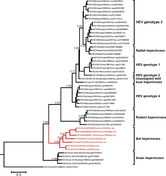
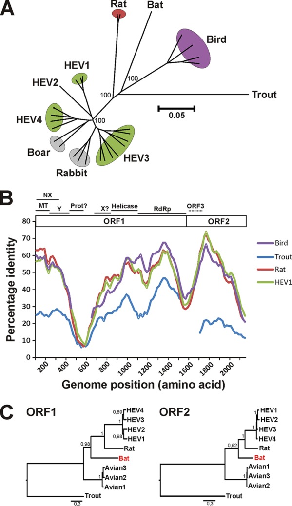
Hepatitis E virus (HEV) is one of the most common causes of acute hepatitis in tropical and temperate climates. Tropical genotypes 1 and 2 are associated with food-borne and waterborne transmission. Zoonotic reservoirs (mainly pigs, wild boar, and deer) are considered for genotypes 3 and 4, which exist in temperate climates. In view of the association of several zoonotic viruses with bats, we analyzed 3,869 bat specimens from 85 different species and from five continents for hepevirus RNA. HEVs were detected in African, Central American, and European bats, forming a novel phylogenetic clade in the family Hepeviridae. Bat hepeviruses were highly diversified and comparable to human HEV in sequence variation. No evidence for the transmission of bat hepeviruses to humans was found in over 90,000 human blood donations and individual patient sera. Full-genome analysis of one representative virus confirmed formal classification within the family Hepeviridae. Sequence- and distance-based taxonomic evaluations suggested that bat hepeviruses constitute a distinct genus within the family Hepeviridae and that at least three other genera comprising human, rodent, and avian hepeviruses can be designated. This may imply that hepeviruses invaded mammalian hosts nonrecently and underwent speciation according to their host restrictions. Human HEV-related viruses in farmed and peridomestic animals might represent secondary acquisitions of human viruses, rather than animal precursors causally involved in the evolution of human HEV.


Figures

Fig 1
Fig 1
Sampling sites and covered bat evolutionary lineages. (A) Sampling sites and numbers of sampled species and specimens per family. (B) Bat evolutionary lineages according to data reported previously (67). Bat families for which samples were tested in this study are shown in boldface type. The names of bat species which tested positive for hepeviruses are shown in red type next to their family designations.
Fig 2
Fig 2
Solid-organ distribution of bat hepevirus BS7 in an Eptesicus serotinus bat. Virus concentrations assessed by strain-specific real-time RT-PCR using quantified in vitro-transcribed RNA controls are given for individual tissue specimens. Since this animal was brought dead to a bat shelter, no blood specimen could be taken due to coagulation.
Fig 3
Fig 3
Partial RdRp gene phylogeny of the family Hepeviridae, including novel bat viruses. The Bayesian phylogeny was generated with MrBayes V3.1 (30), using a general time-reversible (GTR) model with a gamma distribution (G) across sites and a proportion of invariant sites (I) (GTR+G+I) as the substitution model; otherwise default settings were used, and 4,000,000 generations were sampled every 100 steps. In agreement with tree topologies from the full-length ORF1 genes (Fig. 5C), a monophyly prior was set on the root of all mammalian hepeviruses in order to stabilize the phylogenetic reconstruction over this shorter sequence fragment. After an exclusion of 15,000 of the total 40,000 trees, the final tree was annotated and visualized with TreeAnnotator and FigTree from the BEAST package. Values at the nodes indicate the fraction of times that each node was represented within the 95% highest posterior density interval of the trees. Values below 0.7 and those overlapping with taxon names are hidden for clarity of presentation. Branches leading to novel bat viruses and the corresponding taxon names are shown in red. The scale bar indicates genetic distance. The partial RNA-dependent RNA polymerase (RdRp) alignment comprised 324 nucleotides corresponding to positions 4,282 to 4,605 in an HEV genotype 1 prototype strain (GenBank accession number AF459438).
Fig 4
Fig 4
Serologic testing of bat sera for antibodies to human HEV with an HEV-specific indirect immunofluorescence assay. Slides carrying human embryonic kidney 293T cells transiently expressing the full-length ORF2 protein from a human HEV genotype 1 strain were incubated with bat sera (diluted 1:40) from eight different species. To allow the evaluation of the reaction specificity, the transfection efficiency was optimized to yield only 5 to 10% of cells expressing HEV antigen. One HEV RT-PCR-positive species (Vampyrodes caraccioli [PB10/445]) and three different RT-PCR-negative species (Hipposideros gigas [GB557], Rousettus aegyptiacus [GB159], and Miniopterus inflatus [GB475]) are shown. Notably, R. aegyptiacus specimen GB159 was chosen because we realized that it reacted nonspecifically with all cells, including those not expressing HEV antigen, and we wanted to demonstrate its clear discrimination from seropositive human sera. Detection was done by incubation with goat anti-bat immunoglobulin (Ig), followed by donkey anti-goat Ig labeled with cyanine 2. As a control, an anonymous human serum sample from a patient infected with HEV was applied in dilutions of 1:40 and 1:80 (to reduce the background signal). White arrows indicate specific serologic reactivity with HEV ORF2-expressing cells. The bar represents 25 μm. All pictures were taken with identical microscope and camera settings.
Fig 5
Fig 5
Complete genome nucleotide phylogeny, amino acid sequence identity, and ORF1/ORF2 amino acid phylogeny of bat hepevirus BS7 and prototype hepeviruses. (A) Neighbor-joining phylogeny of the complete genomes of members of the Hepeviridae using the nucleotide percentage distance substitution matrix and complete deletion option in MEGA5. Values at deep node points indicate support from 1,000 bootstrap reiterations; those at apical nodes are hidden for clarity of presentation. (B) Amino acid identity plot. The complete ORF1 and ORF2 were translated, concatenated, and compared to avian, rodent, human, and trout prototype hepeviruses. Positions containing gaps in the bat hepevirus were stripped from the alignment. The uncorrected amino acid identity was plotted with a sliding window size of 200 and a step size of 20 amino acids. For orientation, a schematic representation ORF1 and ORF2 is shown with putative nonstructural functional domains as approximated by BLAST comparisons with GenBank reference sequences depicted at the top (MT, methyltransferase; NX, putative ORF NX; Y, Y-like domain; Prot, papain-like cysteine protease; X, X domain/ADP-ribose-binding module; RdRp, RNA-dependent RNA polymerase). The protease and X domains could not be unambiguously identified and are therefore given with question marks. ORF3 is shown with a dotted line, since it is translated in a different reading frame than ORF2 and is shown only for an indication of its genomic position. (C) Bayesian phylogeny of the complete ORF1 and ORF2. Inference of Bayesian phylogenies was done by using MrBayes V3.1 with a WAG amino acid model and 4,000,000 generations sampled every 100 steps. After the exclusion of 10,000 trees as a burn-in, 15,000 final trees were annotated and visualized with TreeAnnotator and FigTree from the BEAST package. Values at the node points indicate posterior probability support (scale bar, genetic distance). GenBank accession numbers for taxa are AF459438 (HEV genotype 1), M74506 (HEV genotype 2), AB301710 (HEV genotype 3), AB220974 (HEV genotype 4), GU345042 (rat hepevirus), AM943647 (avian hepevirus genotype 1), EF206691 (avian hepevirus genotype 2), GU954430 (avian hepevirus genotype 3), and NC_015521 (trout HEV).
Fig 6
Fig 6
Distribution of Hepeviridae partial RdRp and full ORF1 and ORF2 pairwise amino acid distances. Uncorrected pairwise amino acid distances were calculated between members of the family Hepeviridae in the same 108-amino-acid RNA-dependent RNA polymerase (RdRp) alignment as that used for Fig. 3 (A) and in the complete ORF1 (B) and ORF2 (C). The y axis indicates the number of pairwise identity scores within each range represented on the x axis. The bold line indicates a distance cutoff that separates intratypic and intertypic distances within HEV genotypes 1 to 4. The dotted lines indicate a range of possible sequence cutoffs between sequence distances within and between the four suggested hepevirus genera. Distances within NM bat hepeviruses are indicated in light gray. Distances between BS7 bat virus and NM viruses are shown in gray. Distances between PAN926, G19E36, and other bat viruses are shown in black.

Similar articles

Cited by

References

    1. Adjei AA, et al. 2009. Hepatitis E virus infection is highly prevalent among pregnant women in Accra, Ghana. Virol. J. 6:108. - PMC - PubMed
    1. Adlhoch C, Kaiser M, Pauli G, Koch J, Meisel H. 2009. Indigenous hepatitis E virus infection of a plasma donor in Germany. Vox Sang. 97:303–308 - PubMed
    1. Batts W, Yun S, Hedrick R, Winton J. 2011. A novel member of the family Hepeviridae from cutthroat trout (Oncorhynchus clarkii). Virus Res. 158:116–123 - PubMed
    1. Baylis SA, Gartner T, Nick S, Ovemyr J, Blumel J. 2012. Occurrence of hepatitis E virus RNA in plasma donations from Sweden, Germany and the United States. Vox Sang. 103:89–90 - PubMed
    1. Baylis SA, Koc O, Nick S, Blumel J. 2012. Widespread distribution of hepatitis E virus in plasma fractionation pools. Vox Sang. 102:182–183 - PubMed

Publication types

MeSH terms

LinkOut - more resources