. 2012 Sep;86(17):9175-87.
doi: 10.1128/JVI.00309-12. Epub 2012 Jun 13.

The "Bridge" in the Epstein-Barr virus alkaline exonuclease protein BGLF5 contributes to shutoff activity during productive infection

Affiliations

The "Bridge" in the Epstein-Barr virus alkaline exonuclease protein BGLF5 contributes to shutoff activity during productive infection

Daniëlle Horst et al. J Virol. 2012 Sep.

Abstract

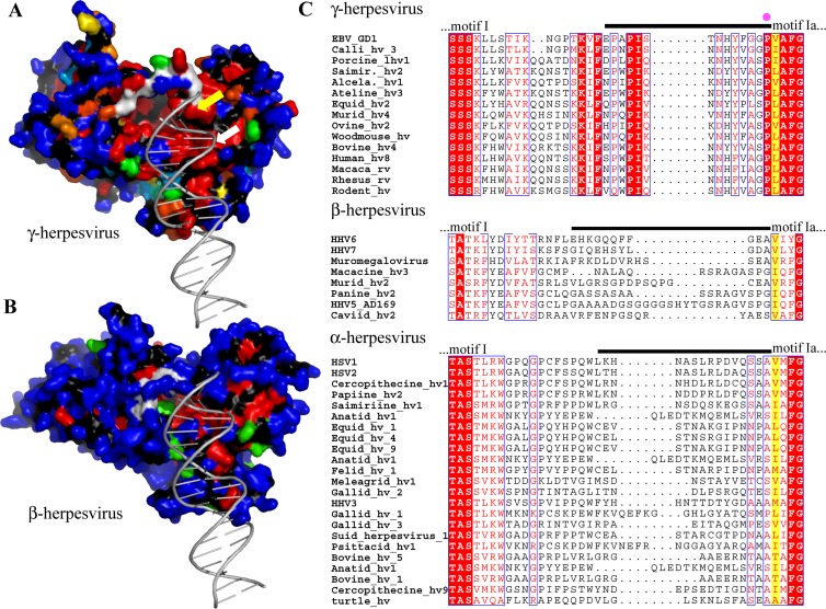
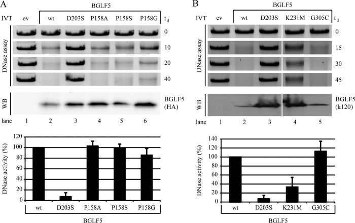
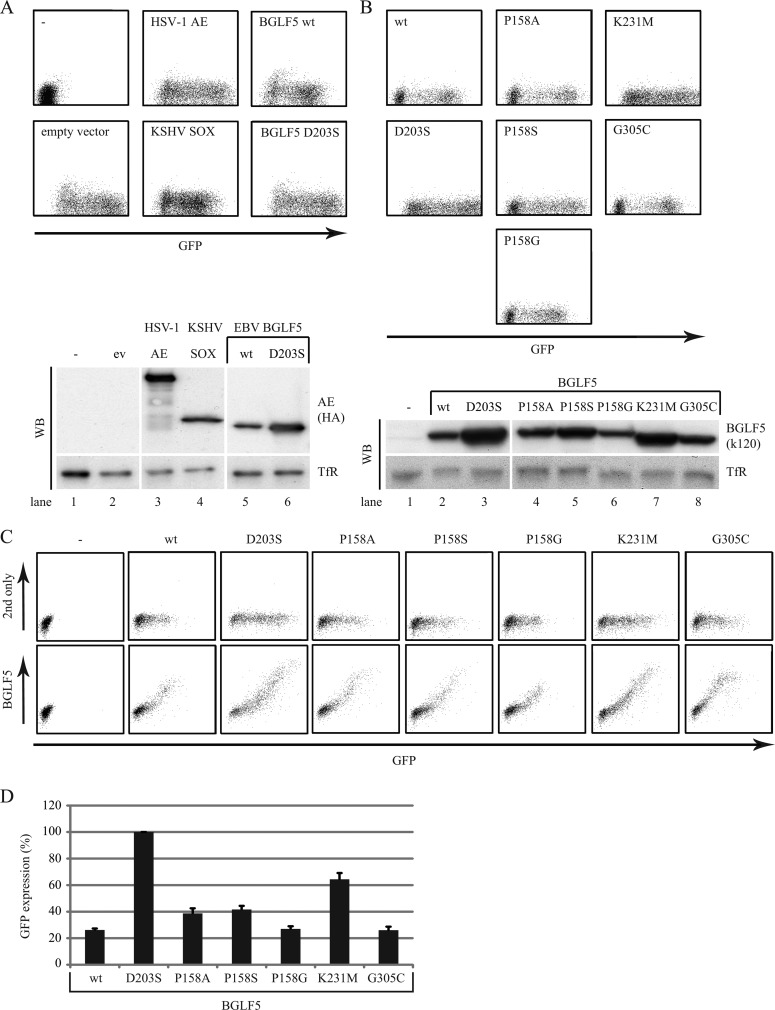
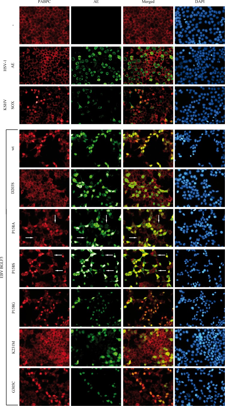
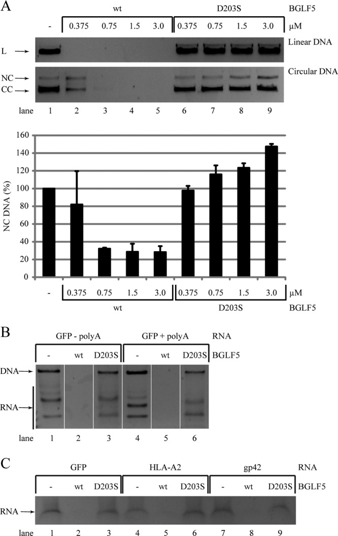
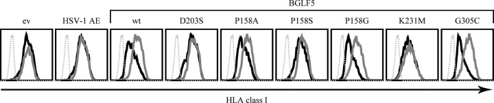
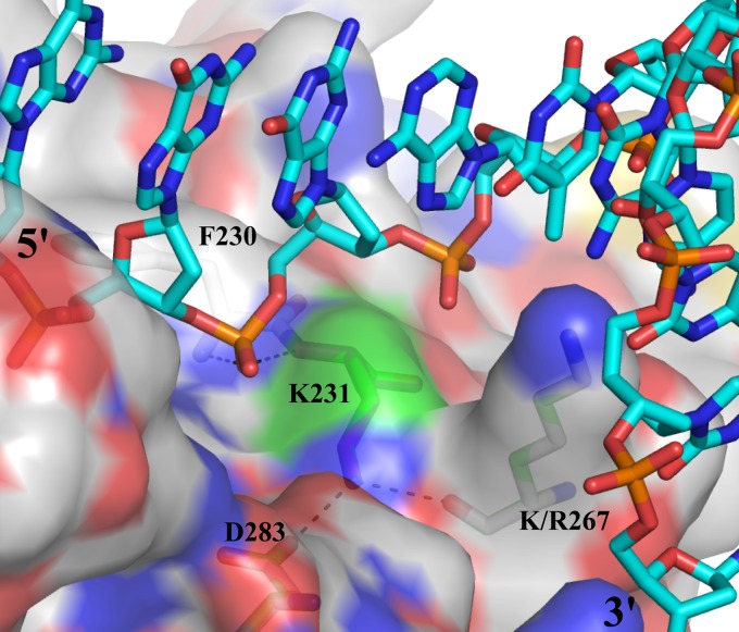
Replication of the human herpesvirus Epstein-Barr virus drastically impairs cellular protein synthesis. This shutoff phenotype results from mRNA degradation upon expression of the early lytic-phase protein BGLF5. Interestingly, BGLF5 is the viral DNase, or alkaline exonuclease, homologues of which are present throughout the herpesvirus family. During productive infection, this DNase is essential for processing and packaging of the viral genome. In contrast to this widely conserved DNase activity, shutoff is only mediated by the alkaline exonucleases of the subfamily of gammaherpesviruses. Here, we show that BGLF5 can degrade mRNAs of both cellular and viral origin, irrespective of polyadenylation. Furthermore, shutoff by BGLF5 induces nuclear relocalization of the cytosolic poly(A) binding protein. Guided by the recently resolved BGLF5 structure, mutants were generated and analyzed for functional consequences on DNase and shutoff activities. On the one hand, a point mutation destroying DNase activity also blocks RNase function, implying that both activities share a catalytic site. On the other hand, other mutations are more selective, having a more pronounced effect on either DNA degradation or shutoff. The latter results are indicative of an oligonucleotide-binding site that is partially shared by DNA and RNA. For this, the flexible "bridge" that crosses the active-site canyon of BGLF5 appears to contribute to the interaction with RNA substrates. These findings extend our understanding of the molecular basis for the shutoff function of BGLF5 that is conserved in gammaherpesviruses but not in alpha- and betaherpesviruses.


Figures

Fig 1
Fig 1
Structure and amino acid sequence comparisons for herpesvirus exonucleases. (A) Sequence conservation among 28 AE sequences of gammaherpesviruses (see Table 1) mapped onto the surface of the EBV nuclease BGLF5. Conservation of the side chains decreases from red (strictly conserved) over orange, yellow and green to blue (variable residues). Main chain atoms are colored in black, the ones of the bridge are colored in white. The position of a dsDNA substrate is derived from the KSHV dsDNA-SOX complex (2). The position of V159, the equivalent of a residue involved in the processivity of lambda exonuclease (54), is indicated by a yellow arrow, the white arrow indicates the K231 residue mutated in the present study. (B) Analysis of the sequence conservation within 8 betaherpesvirus exonucleases (see Table 1) based on a theoretical model of their structure. Colors are as defined in panel A. (C) Sequence conservation in the region of the bridge, which is indicated with a black line above the amino acid sequence alignments. A yellow background marks the residues corresponding to the amino acid essential for exonuclease activity in lambda exonuclease (54). The pink dot marks the position of EBV P158. hv, herpesvirus; rv, rhadinovirus.
Fig 2
Fig 2
Location of the studied mutations within the EBV BGLF5 protein. Within the structural model of the EBV nuclease, the amino acid substitutions under study are colored as follows: P158 mutants in dark blue, D203S mutant in purple, K231M mutant in yellow, and G305C mutant in red. The exact context and conservation of the mutations is shown in Fig. S1 in the supplemental material. The flexible bridge is indicated by an arrow. (PDB entry 2W4B.)
Fig 3
Fig 3
Point mutations within EBV BGLF5 differentially affect DNase activity. (A and B) Linearized pcDNA3 DNA was incubated with the indicated (mutant) BGLF5 proteins synthesized as in vitro translation products. Samples of pcDNA3 substrate taken before and at the indicated time points during the degradation reaction (td, time of degradation in minutes) were resolved by agarose gel electrophoresis and visualized by ethidium bromide staining. To visualize the AE proteins as added to the enzyme reaction mixtures, 5-μl portions of the in vitro translation products were separated by SDS-PAGE and analyzed by Western blotting (WB) using either antibodies specific for the HA tag added to the C terminus of BGLF5 (3F10) (A) or with a BGLF5-specific polyclonal rabbit serum (k120) (B). For quantification of the DNase assays, the DNA amounts present after the degradation reaction were corrected for the corresponding empty vector control samples. Subsequently, the DNase activity of the BGLF5 proteins was determined with the activity of wild-type BGLF5 set at 100%. The standard deviations are represented by the error bars. All proteins were analyzed in at least four independent experiments, except for the K231M and G305C proteins that were analyzed in duplicate. ev, empty vector; wt, wild type.
Fig 4
Fig 4
Point mutations within BGLF5 differentially affect shutoff activity. (A and B) 293T cells were cotransfected with the reporter plasmid pcDNA3-IRES-nlsGFP and a pcDEF vector encoding HSV-1 AE, KSHV SOX, wild-type EBV BGLF5 (wt), or the indicated BGLF5 mutants (at a 1:4 ratio). At 48 h after transfection, the GFP intensity was analyzed by flow cytometry as a measure of the shutoff activity. Cellular expression of the various AE proteins was assessed by Western blot analysis with antibodies specific for the HA tag (3F10) (A) or BGLF5 (k120) (B). Transferrin receptor (TfR)-specific MAb H68.4 was taken along as a loading control. For the flow cytometric data, the results of one representative experiment out of five independent experiments are shown. For the Western blots, the results of one representative experiment out of two independent experiments are shown. (C) 293T cells were transfected with a pcDNA3-IRES-nlsGFP vector encoding wild-type BGLF5 (wt) or the indicated BGLF5 mutants. Nontransfected cells were taken along as a control. At 48 h after transfection, the cells were fixed, permeabilized, and stained for intracellular expression of BGLF5 (MAb 311H) and analyzed by flow cytometry. The results of one representative experiment out of two independent experiments are shown. (D) Quantification of flow cytometric data. The geometric mean fluorescence intensities for GFP within the BGLF5-positive cells depicted in panel C was determined. Subsequently, the values were set at 100% for the BGLF5 D203S catalytic site mutant. The standard deviations are represented by the error bars. All samples were analyzed in duplicate.
Fig 5
Fig 5
EBV BGLF5 induces nuclear relocalization of PABPC, an activity that is correlated with shutoff. 293T cells were transfected with pcDEF plasmids encoding the individual herpesvirus AE proteins as in Fig. 4A and B. After 24 h of transfection, the cells were subjected to triple-label immunofluorescence analysis: PABPC expression was visualized by PABPC-specific antibodies (MAb 10E10 and anti-mouse-Cy3; left panels), AE expression was visualized with HA-specific antibodies (MAb 3F10 and anti-rat Alexa 488; second panels for negative control, HSV-1 AE and KSHV SOX samples) or with BGLF5-specific antibodies (k120 polyclonal antisera and anti-rabbit Alexa 488; second panels for [mutant] BGLF5 samples), and cells were counterstained with DAPI to identify nuclei (right panels). The white arrows indicate cells with low levels of the P158A and P158S mutant proteins. The KSHV SOX, BGLF5 wt, and D203S proteins were analyzed in triplicate; the other proteins were analyzed in duplicate.
Fig 6
Fig 6
Specificities of EBV BGLF5-mediated DNA and RNA degradation. (A) To assess DNase activity, linear and circular pMaxGFP plasmids (0.05 pmol of DNA) were incubated with increasing concentrations of wt or D203S mutant EBV BGLF5 recombinant proteins in the presence of 5 mM Mg2+. After 2 h at 37°C, samples were resolved by agarose gel electrophoresis and visualized by ethidium bromide staining. The results of one representative experiment out of four independent experiments are shown. For quantification of the DNase assays, the NC DNA amounts present after the degradation reaction were corrected for the control samples (set at 100%). The standard deviations are represented by error bars. The quantification is based on two replicate experiments. L, linear DNA; CC, closed circle; NC, nicked circle. (B and C) The specificity of the RNase activity was studied by incubating wt or D203S mutant EBV BGLF5 recombinant proteins with in vitro-transcribed RNA in the presence of 10 mM Mn2+. (B) GFP RNA was synthesized from a plasmid linearized before (−polyA) or after (+polyA) the polyadenylation consensus sequence. (C) RNAs for a foreign protein (GFP), a cellular protein (HLA-A2), or a viral protein (EBV gp42) were used. After degradation periods of 30 min (B) or 6 h (C) at 37°C, the samples were resolved by agarose gel electrophoresis and visualized by ethidium bromide staining. DNA and RNA bands were identified according to their susceptibility to digestion with an RNase A/T1 mix (Ambion) (data not shown). Using this approach, the GFP (+polyA) and cellular mRNAs were analyzed once, the GFP (−polyA) and viral mRNAs were analyzed in at least two independent experiments.
Fig 7
Fig 7
HLA class I downregulation by BGLF5 mutants depends on shutoff activity. 293T cells were transfected with a pcDNA3-IRES-nlsGFP vector encoding HSV-1 AE, wild-type EBV BGLF5, or the indicated BGLF5 mutants. As a control, the empty vector was transfected. At 48 h after transfection, the cells were stained for cell surface expression of HLA class I (MAb W6/32) and analyzed by flow cytometry. Dashed gray line, secondary antibody only; solid gray line, GFP-negative cells; solid black line, GFP-positive cells. The results of one representative experiment out of two independent experiments are shown. ev, empty vector; wt, wild type.
Fig 8
Fig 8
Role of the EBV BGLF5 residue K231. View of the KSHV SOX structure in complex with dsDNA (2). Only one of the DNA strands is shown (cyan carbon atoms). The protein (white carbon atoms) is represented with a semitransparent surface with some key residues indicated. Residue K231 is shown with green carbon atoms. Residues are labeled with the numbers corresponding to EBV BGLF5. These residues are conserved between BGLF5 and SOX with the exception of an R/K substitution. Dotted lines show hydrogen bonds.

References

    1. Altschul SF, et al. 1997. Gapped BLAST and PSI-BLAST: a new generation of protein database search programs. Nucleic Acids Res. 25:3389–3402 - PMC - PubMed
    1. Bagneris C, Briggs LC, Savva R, Ebrahimi B, Barrett TE. 2011. Crystal structure of a KSHV-SOX-DNA complex: insights into the molecular mechanisms underlying DNase activity and host shutoff. Nucleic Acids Res. 39:5744–5756 - PMC - PubMed
    1. Barnstable CJ, et al. 1978. Production of monoclonal antibodies to group A erythrocytes, HLA and other human cell surface antigens-new tools for genetic analysis. Cell 14:9–20 - PubMed
    1. Benkert P, Biasini M, Schwede T. 2011. Toward the estimation of the absolute quality of individual protein structure models. Bioinformatics 27:343–350 - PMC - PubMed
    1. Buisson M, et al. 2009. A bridge crosses the active-site canyon of the Epstein-Barr virus nuclease with DNase and RNase activities. J. Mol. Biol. 391:717–728 - PubMed

Publication types

MeSH terms

Substances

LinkOut - more resources