

Review
doi: 10.1136/bcr.03.2011.4022.
Jejunal cavernous lymphangioma
Affiliations
- PMID: 22696733
- PMCID: PMC3094793
- DOI: 10.1136/bcr.03.2011.4022
Item in Clipboard
Review
Jejunal cavernous lymphangioma
BMJ Case Rep.
.
Abstract
Cavernous lymphangiomas are usually identified in infants and children with the majority of lesions found around the head and neck, trunk or extremities. Tumours affecting the intra-abdominal organs are rare. The authors report a case of small bowel cavernous lymphangioma arising within the jejunum of a 34-year-old woman presenting with dyspnoea and anaemia, and review the existing literature relating to this uncommon tumour.
Conflict of interest statement
Figures
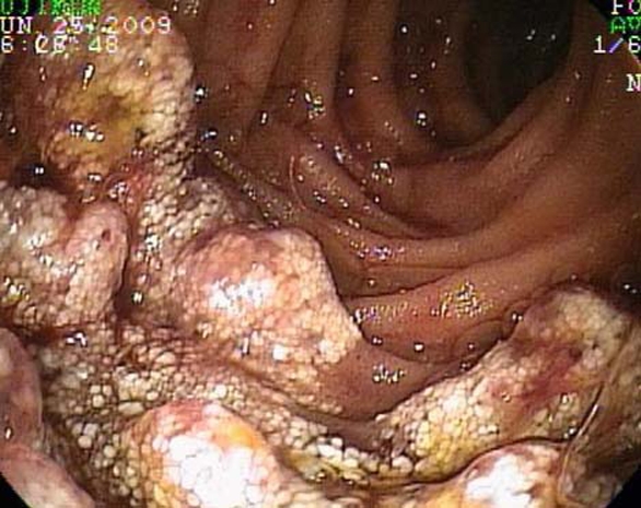
Enteroscopic appearance of the lymphangioma.
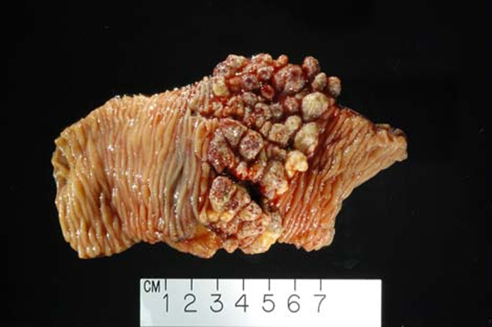
This gross photograph of the segmental jejunal resection illustrates the striking multinodular and polypoid appearance of the jejunal lymphangioma, along with the entirely normal appearing adjacent jejunum.
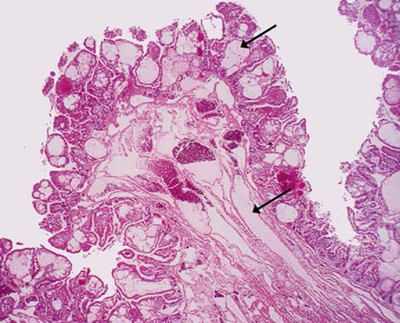
The mucosa and submucosa are expanded and replaced by innumerable dilated lymphatics (arrows).
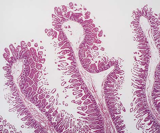
The jejunum adjacent to the lymphangioma was normal histologically, as illustrated here with intact villous architecture, mucosa and submucosa, lacking dilated lymphatic spaces. This feature excludes diffused lymphangiomatosis from the differential.
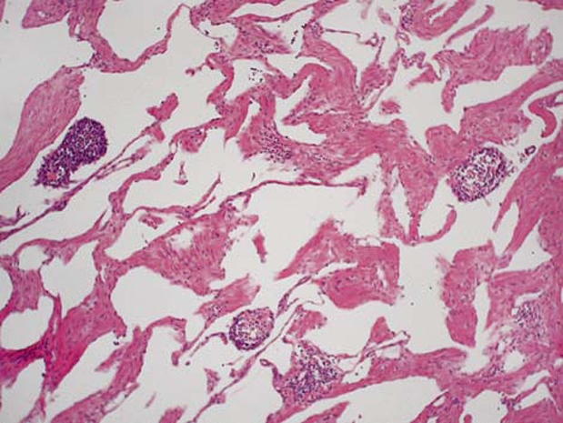
In addition to the mass forming lymphangioma occupying the wall of the jejunum, an additional focal mesenteric lymphangioma was identified in the resection specimen. The fibromuscular walls making up the lymphatic vessels and luminal benign lymphoid aggregates are illustrated.
References
-
- Alqahtani A, Nguyen LT, Flageole H, et al. 25 years’ experience with lymphangiomas in children. J Pediatr Surg 1999;34:1164–8 - PubMed
-
- Fonkalsrud EW. Congenital malformations of the lymphatic system. Semin Pediatr Surg 1994;3:62–9 - PubMed
-
- Hanagiri T, Baba M, Shimabukuro T, et al. Lymphangioma in the small intestine: report of a case and review of the Japanese literature. Surg Today 1992;22:363–7 - PubMed
-
- Rieker RJ, Quentmeier A, Weiss C, et al. Cystic lymphangioma of the small-bowel mesentery: case report and a review of the literature. Pathol Oncol Res 2000;6:146–8 - PubMed
-
- Uncu H, Erdem E, Kuterdem E. Lymphangiomas of the ileum: a report of two cases and a review of the literature. Surg Today 1997;27:542–5 - PubMed
Publication types
MeSH terms
LinkOut - more resources
Full Text Sources
Medical