Review
. 2012 Jun 14:9:20.
doi: 10.1186/1743-8977-9-20.

Autophagy and lysosomal dysfunction as emerging mechanisms of nanomaterial toxicity

Affiliations
Review

Autophagy and lysosomal dysfunction as emerging mechanisms of nanomaterial toxicity

Stephan T Stern et al. Part Fibre Toxicol. .

Abstract

The study of the potential risks associated with the manufacture, use, and disposal of nanoscale materials, and their mechanisms of toxicity, is important for the continued advancement of nanotechnology. Currently, the most widely accepted paradigms of nanomaterial toxicity are oxidative stress and inflammation, but the underlying mechanisms are poorly defined. This review will highlight the significance of autophagy and lysosomal dysfunction as emerging mechanisms of nanomaterial toxicity. Most endocytic routes of nanomaterial cell uptake converge upon the lysosome, making the lysosomal compartment the most common intracellular site of nanoparticle sequestration and degradation. In addition to the endo-lysosomal pathway, recent evidence suggests that some nanomaterials can also induce autophagy. Among the many physiological functions, the lysosome, by way of the autophagy (macroautophagy) pathway, degrades intracellular pathogens, and damaged organelles and proteins. Thus, autophagy induction by nanoparticles may be an attempt to degrade what is perceived by the cell as foreign or aberrant. While the autophagy and endo-lysosomal pathways have the potential to influence the disposition of nanomaterials, there is also a growing body of literature suggesting that biopersistent nanomaterials can, in turn, negatively impact these pathways. Indeed, there is ample evidence that biopersistent nanomaterials can cause autophagy and lysosomal dysfunctions resulting in toxicological consequences.


Figures

Figure 1
Figure 1
An overview of phagocytic and non-phagocytic pathways. A) Phagocytosis occurs in macrophages through an actin-based mechanism involving interaction with various specialized cell surface receptors (e.g., mannose, IgG, complement, Fcγ receptors). The foreign particles recognized by specific receptors, often targeting surface-bound opsonins, are internalized to form endocytic vesicles called phagosomes. The fusion of phagosomes with the lysosomal compartment leads to the formation of phagolysosomes, where the foreign particles are enzymatically degraded. B) Clathrin-mediated endocytosis involves the formation of vesicles from triskelion clathrin-coated regions of the plasma membrane. The triskelion clathrin in the cytosol are then recycled back to the plasma membrane followed by movement of ingested materials from early endosome to the late endosome, finally fusing with lysosome to form the lysosome-endosome hybrid. The materials are then degraded by the low pH and enzyme-rich environment of the endo-lysosomal vesicle. C) Caveolin-mediated endocytosis involves internalization through caveolin (a dimeric protein) enriched invaginations. The cytosolic caveolin vesicle then delivers its contents to endosomes, to form caveosomes which can avoid lysosomal enzymatic degradation, and are transported along the cytoskeleton to the endoplasmic reticulum/golgi complex. D) Macropinocytosis is a clathrin- and caveolin-independent pathway. It involves the formation of large vesicles called macropinosomes, which occurs through actin filament driven plasma membrane protrusions. The contents are degraded following fusion with the lysosomal compartment. (Figure partially adapted from Hillaireau and Couvreur, Cell. Mol. Life Sci. 2009, 66, 2873–2896).
Figure 2
Figure 2
Autophagy. 1) During autophagy, a double layer membrane, the autophagosome, is formed that surrounds proteins and damaged organelles destined for degradation. 2) The autophagosome then merges with the lysosome, where hydrolytic enzymes in the lysosome dismantle the autophagosome contents. 3) The autophagy pathway is interconnected with the endocytosis pathways, with most endosomes eventually merging with the lysosome.
Figure 3
Figure 3
Mechanisms of autophagy and lysosomal dysfunction toxicity. The initiators of autophagy and lysosomal dysfunction toxicity, displayed in light blue text in the figure, include blockade of vesicle trafficking, lysosomal membrane permeabilization (LMP), and autophagy dysregulation. Nanoparticles could potentially cause autophagy dysfunction by overloading or directly damaging the lysosomal compartment, or altering the cell cytoskeleton, resulting in blockade of autophagosome-lysosome fusion. Nanoparticles could also directly affect lysosomal stability by inducing lysosomal oxidative stress, alkalization, osmotic swelling, or causing detergent-like disruption of the lysosomal membrane itself, resulting in LMP. Toxic effectors (lysosomal iron, cytosolic acidification, hydrolytic enzymes, reactive oxygen species, and the NLRP3 inflammasome) are displayed in dark blue. Conditions resulting from effector-mediated loss of homeostasis (oxidative stress, inflammation, ER stress, disrupted mitophagy, accumulation of ubiquitinated protein aggregates, and mitochondrial perturbation) are displayed in green. Finally, this loss of homeostasis can result in the cell death pathways necrosis, and Apoptotic (type I) and autophagic (type II) cell death; displayed in red (see text for details).
Figure 4
Figure 4
Nanoparticle-induced autophagy. A) Recent evidence supporting ubiquitination of nanomaterials directly, or indirectly through colocalization with protein aggregates, suggests that cells may select nanomaterials for autophagy through a p62-LC3 II pathway similar to invading pathogens (see text). B) Data also supports nanomaterial-induced alteration of autophagy signaling pathways, including: 1) induction of oxidative stress-dependent signaling (e.g., ER stress, mitochondrial damage), 2) suppression of Akt-mTOR signaling, and 3) alteration of autophagy related gene/protein expression.
Figure 5
Figure 5
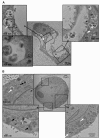
Transmission electron micrographs detailing autophagic vacuoles. Porcine kidney cells (LLC-PK1) were treated for 24 h with either (A) 10 nM CdSe or (B) 100 nM InGaP quantum dots. The white arrows indicate autophagic vacuoles containing cellular debris. The black arrows indicate lysosomal remnants, consisting of multilamellar vacuoles and electron-dense deposits. (Stern ST, et al, Induction of Autophagy in Porcine Kidney Cells by Quantum Dots: A Common Cellular Response to Nanomaterials? Tox. Sci. 2008, 106(1), 140–152, by permission of Oxford University Press.)

Similar articles

Cited by

References

    1. Li N, Xia T, Nel AE. The role of oxidative stress in ambient particulate matter-induced lung diseases and its implications in the toxicity of engineered nanoparticles. Free Radic Biol Med. 2008;44:1689–1699. doi: 10.1016/j.freeradbiomed.2008.01.028. - DOI - PMC - PubMed
    1. Stern ST, Johnson DN. Role for nanomaterial-autophagy interaction in neurodegenerative disease. Autophagy. 2008;4:1097–1100. - PubMed
    1. Klionsky DJ. Autophagy: from phenomenology to molecular understanding in less than a decade. Nat Rev Mol Cell Biol. 2007;8:931–937. doi: 10.1038/nrm2245. - DOI - PubMed
    1. De Duve C, De Barsy T, Poole B, Trouet A, Tulkens P, Van Hoof F. Commentary. Lysosomotropic agents. Biochem Pharmacol. 1974;23:2495–2531. doi: 10.1016/0006-2952(74)90174-9. - DOI - PubMed
    1. Hillaireau H, Couvreur P. Nanocarriers' entry into the cell: relevance to drug delivery. Cell Mol Life Sci. 2009;66:2873–2896. doi: 10.1007/s00018-009-0053-z. - DOI - PMC - PubMed

Publication types

LinkOut - more resources