

Case Reports
doi: 10.1186/2047-783X-17-16.
Vascular diaphragmatic hernia in a patient with cirrhosis first case report
Affiliations
- PMID: 22697282
- PMCID: PMC3439304
- DOI: 10.1186/2047-783X-17-16
Item in Clipboard
Case Reports
Vascular diaphragmatic hernia in a patient with cirrhosis first case report
Eur J Med Res.
.
Abstract
We report the case of an adult patient recently diagnosed with cirrhosis. The ultrasound evaluation described a multinodular inhomogeneous liver, requiring a magnetic resonance imaging scan for further characterization. The performed magnetic resonance imaging examination confirmed the diagnosis of cirrhosis associated with portal hypertension and detected a vascular left transdiaphragmatic hernia. Although various types of diaphragmatic hernias have been described - congenital or acquired - to the best of our knowledge, this type of pathology has never been reported.
Figures

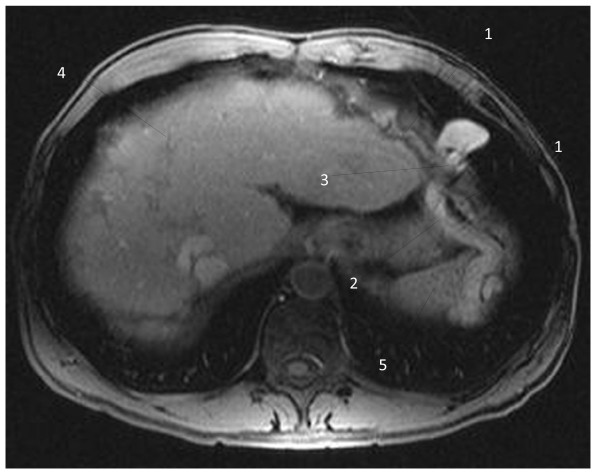
T1-weighted magnetic resonance image of the vascular diaphragmatic hernia (non-contrast image), axial view. 1 - Left diaphragm muscle; 2 - descending part of the collateral aberrant vein; 3 - diaphragmatic defect corresponding to the base of the vascular loop; 4 - liver; 5 - spleen.
T1-weighted magnetic resonance image of the vascular diaphragmatic hernia (after contrast administration), axial view. 1 - Left diaphragm muscle; 2 - descending part of the collateral aberrant vein; 3 - diaphragmatic defect corresponding to the base of the vascular loop; 4 - liver; 5 - spleen.

T1-weighted magnetic resonance image of the vascular diaphragmatic hernia (after contrast administration), coronal view. 1 - Left diaphragm muscle; 2 - thoracic part of the herniated vein; 3 - recanalized umbilical vein; 4 – liver.

T1-weighted magnetic resonance image of the vascular diaphragmatic hernia (after contrast administration), coronal view. 1 - Left diaphragm muscle; 2 - abdominal part of the herniated vein; 3 - diaphragmatic defect corresponding to the base of the vascular loop; 4 - liver.
References
-
- Langham MR, Kays DW, Ledbetter DJ, Frentzen B, Sanford LL, Richards DS. Congenital diaphragmatic hernia: epidemiology and outcome. Clin Perinatol. 1996;23:671–688. - PubMed
-
- Sarin SK, Govil A, Jain AK, Guptan RC, Issar SK, Jain M, Murthy NS. Prospective randomized trial of endoscopic sclerotherapy versus variceal band ligation for esophageal varices: influence on gastropathy, gastric varices and variceal recurrence. J Hepatol. 1997;26:826–832. doi: 10.1016/S0168-8278(97)80248-6. - DOI - PubMed
Publication types
MeSH terms
LinkOut - more resources
Full Text Sources
Medical
