. 2012 Nov;16(11):2736-44.
doi: 10.1111/j.1582-4934.2012.01596.x.

Fluoxetine induces vasodilatation of cerebral arterioles by co-modulating NO/muscarinic signalling

Affiliations

Fluoxetine induces vasodilatation of cerebral arterioles by co-modulating NO/muscarinic signalling

Keren Ofek et al. J Cell Mol Med. 2012 Nov.

Abstract

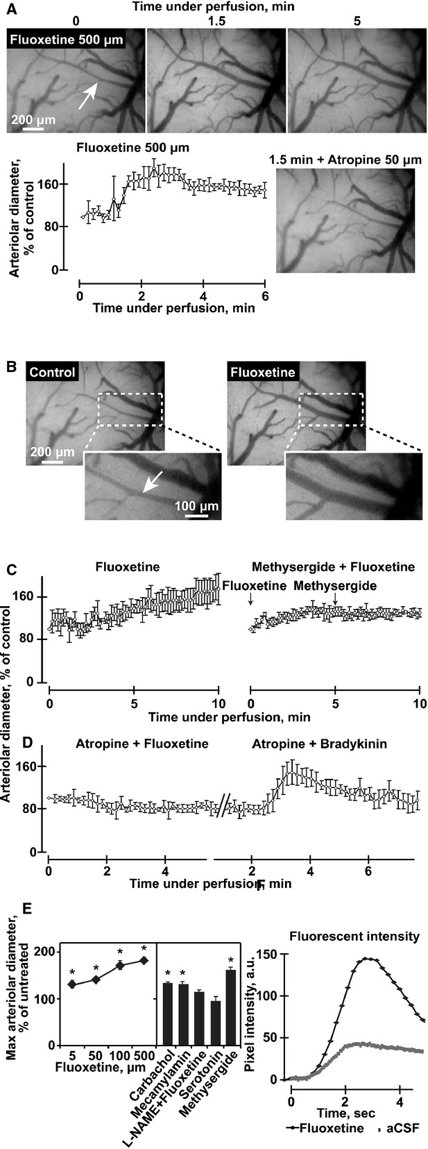
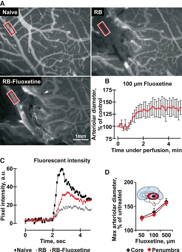
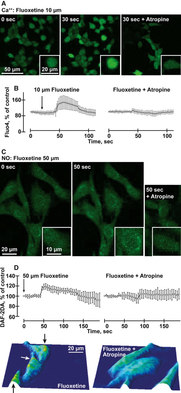
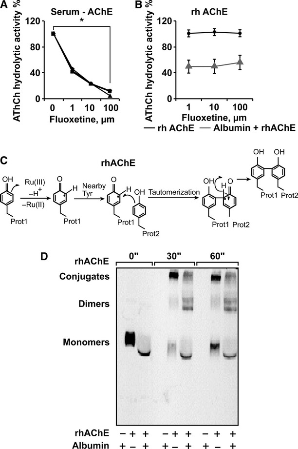
Ischaemic stroke patients treated with Selective Serotonin Reuptake Inhibitors (SSRI) show improved motor, cognitive and executive functions, but the underlying mechanism(s) are incompletely understood. Here, we report that cerebral arterioles in the rat brain superfused with therapeutically effective doses of the SSRI fluoxetine showed consistent, dose-dependent vasodilatation (by 1.2 to 1.6-fold), suppressible by muscarinic and nitric oxide synthase (NOS) antagonists [atropine, NG-nitro-l-arginine methyl ester (l-NAME)] but resistant to nicotinic and serotoninergic antagonists (mecamylamine, methylsergide). Fluoxetine administered 10-30 min. following experimental vascular photo-thrombosis increased arterial diameter (1.3-1.6), inducing partial, but lasting reperfusion of the ischaemic brain. In brain endothelial b.End.3 cells, fluoxetine induced rapid muscarinic receptor-dependent increases in intracellular [Ca(2+) ] and promoted albumin- and eNOS-dependent nitric oxide (NO) production and HSP90 interaction. In vitro, fluoxetine suppressed recombinant human acetylcholinesterase (rhAChE) activity only in the presence of albumin. That fluoxetine induces vasodilatation of cerebral arterioles suggests co-promotion of endothelial muscarinic and nitric oxide signalling, facilitated by albumin-dependent inhibition of serum AChE.


Figures

Fig. 1
Fig. 1
Fluoxetine induces a rapid, robust and long-lasting muscarinic/eNOS-mediated vasodilatation. (A) Representative images of pial arterioles in the rat cerebral cortex. Shown are photographs of the cranial windows at the noted time-points in minutes and administered pharmakons. Note rapid response and inhibition of 500 μM fluoxetine-mediated vasodilatation inhibition by 50 μM atropine. Arterioles and venules were identified visually and arterioles identity confirmed by their earlier filling with the fluorescent dye in the angiography experiments. B. Representative images of pial arterioles in the rat cerebral cortex. Shown are photographs before (Control) and 1.5 min. under 100 μM fluoxetine perfusion (fluoxetine). Below: enlarged dashed frames. As the focus is not similar, we performed diameter analysis for multiple consecutive images acquired at 0.5–1 Hz. (C and D) Arteriolar diameter presented as percentage from control (averages ± S.D. for 5 points along an arteriole) following perfusion of 100 μM fluoxetine without or with 50 μM atropine, 5 μM fluoxetine with 100 μM methysegrid, or 100 μM bradykinin with 50 μM atropine-each presenting one out of four experimental animals with similar outcome, except for one animal in the bradikinin + atropine test (anova P < 0.001). (E) Left: Maximal arterial diameter following perfusion of the noted compounds, presented as average ± S.D. at ± 10 sec. around the peak. The aterioles we investigated had a mean diameter of 24.35 ± 9.72 μm before pharmacological manipulation. Right: Carbachol, mecamylamine and methysergide (50 μM), but not serotonin (50 mM) enlarges arteriolar diameter, and l-NAME (1 mM) blocks the 500 μM fluoxetine-induced enlargement in arteriolar diameter (anova P < 0.001). (F) Arterial fluorescent intensity representing cerebral blood flow, in pixel intensity following bolus injection of fluorescein sodium salt of fluoxetine versus ‘control angiography’. (anova P < 0.001, N = 3).
Fig. 2
Fig. 2
Fluoxetine increases blood flow after laser-induced small artery ischaemia. (A) Representative images of the naïve pial vasculature, before (left) and after photothrombosis (Rose Bengal – RB, right) and after 100 μM fluoxetine treatment (RB + Fluoxetine, below) following i.v. injection of fluorescein sodium salt. Red-framed rectangles indicate areas of blood flow measurement. (B) Arteriolar diameter presented as percentage of control (average ± S.D. for 5 different points within the arteriole) following perfusion of 100 μM fluoxetine in an ischaemic rat. (C) Fluorescent intensity representing cerebral blood flow and the arteriolar peak in the above samples. (anova P < 0.001). (D) Maximal arteriolar diameter following perfusion of fluoxetine in the noted concentrations. Presented are average ± S.D. at 10 sec. around the peak within the core (black) and the penumbra (red). (anova P < 0.001). Inset: Scheme of brain involvement in ischaemic stroke. A core of infarcted tissue (black zone) is surrounded by a peripheral region referred to as the penumbra (red).
Fig. 3
Fig. 3
Fluoxetine induces atropine-suppressible intracellular Ca2+ and NO release in mouse b.End.3 cells (A) Ca++: b.End.3 cells stained with Fluo-4 and treated with 10 μM fluoxetine (FL) for 30 sec. with or without atropine. (B) Quantification of average changes in Ca++ signals in 40 cells under the conditions noted above. (anova P < 0.001). (C) NO: b.End.3 cells stained with DAF-2DA and treated with 50 μM fluoxetine with or without atropine. (D) Quantification of changes in NO signals in 30 cells under the conditions noted above. (anova P < 0.001). Below: Typographical map of the NO signal represents the field of the image above, following fluoxetine treatment of b.End.3 cells with or without atropine. Note high levels near the nucleus (yellow, marked by arrows).
Fig. 4
Fig. 4
Fluoxetine increases the levels of eNOS and its enhancer HSP90 in B.End.3 endothelial cells. (A) Top scheme: Fluoxetine induction of eNOS-HSP90-mediated vasodilatation involves facilitation of NO production by eNOS. (B) Top row: Enhanced HSP90 under fluoxetine. Columns: Quantification (Student's t-test CT-FL P < 0.00). Second row: Enhanced eNOS immunolabeling in b.End.3 cells pretreated for 24 hrs with 10 μM fluoxetine. Columns: Quantification (Student's t-test P < 0.00).Third row: Enhanced HSP90-eNOS colocalizaion under fluoxetine. Columns: Quantification (Student's t-test P < 0.00). Fourth row: DAPI staining showed sustained cell density. (C) B.End.3 cell extract was incubated with anti-eNOS antibodies and precipitates electroblotted. Immunolabeling showed HSP90-induced elevation of eNOS in the fluoxetine-treated cell lysate compared to untreated cells.
Fig. 5
Fig. 5
Fluoxetine reduces rhAChE hydrolytic activity when interacting with albumin (A) AChE activity following 15 min. incubation of human serum samples with the noted μM doses of fluoxetine. Presented are residual percentages of hydrolytic activity from control values of serum samples from three different individuals. Average ± S.D. serum AChE activity without fluoxetine: 146.4 ± 10.8; N = 3. (anova P < 0.001). (B) hrAChE activity following incubation of 15 min. with the noted doses of fluoxetine with and without 70 mg/ml Albumin. (anova P < 0.001). (C) Scheme of photo-induced cross-linking by ruthenium ions. (D) Albumin attenuates the formation of AChE multimeric conjugates in the photo-induced cross-linking assay. Note the photo-induction, within 30 sec. of slowly migrating AChE multimeric conjugates from rhAChE monomers and the attenuated production of these multimers, yielding higher dimer levels in the presence of albumin.

References

    1. Roger VL, Go AS, Lloyd-Jones DM, et al. Heart disease and stroke statistics–2011 update: a report from the American Heart Association. Circulation. 2011;123:e18–209. - PMC - PubMed
    1. Shuaib A, Butcher K, Mohammad AA, et al. Collateral blood vessels in acute ischaemic stroke: a potential therapeutic target. Lancet Neurol. 2011;10:909–21. - PubMed
    1. Li WL, Cai HH, Wang B, et al. Chronic fluoxetine treatment improves ischaemia-induced spatial cognitive deficits through increasing hippocampal neurogenesis after stroke. J Neurosci Res. 2009;87:112–22. - PubMed
    1. Chollet F, Tardy J, Albucher JF, et al. Fluoxetine for motor recovery after acute ischaemic stroke (FLAME): a randomised placebo-controlled trial. Lancet Neurol. 2011;10:123–30. - PubMed
    1. Narushima K, Paradiso S, Moser DJ, et al. Effect of antidepressant therapy on executive function after stroke. Br J Psychiatry. 2007;190:260–5. - PubMed

Publication types

MeSH terms