

Evolution and development of the vertebrate neck
- PMID: 22697305
- PMCID: PMC3552416
- DOI: 10.1111/j.1469-7580.2012.01530.x
Evolution and development of the vertebrate neck
Abstract
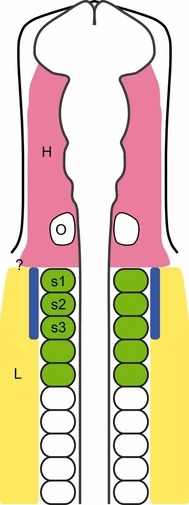
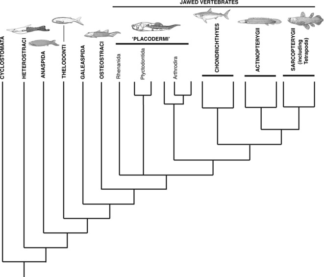
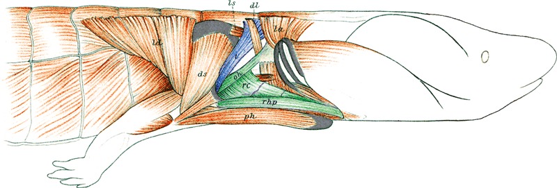
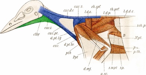
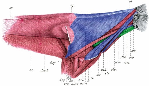
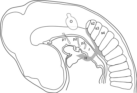
Muscles of the vertebrate neck include the cucullaris and hypobranchials. Although a functional neck first evolved in the lobe-finned fishes (Sarcopterygii) with the separation of the pectoral/shoulder girdle from the skull, the neck muscles themselves have a much earlier origin among the vertebrates. For example, lampreys possess hypobranchial muscles, and may also possess the cucullaris. Recent research in chick has established that these two muscles groups have different origins, the hypobranchial muscles having a somitic origin but the cucullaris muscle deriving from anterior lateral plate mesoderm associated with somites 1-3. Additionally, the cucullaris utilizes genetic pathways more similar to the head than the trunk musculature. Although the latter results are from experiments in the chick, cucullaris homologues occur in a variety of more basal vertebrates such as the sharks and zebrafish. Data are urgently needed from these taxa to determine whether the cucullaris in these groups also derives from lateral plate mesoderm or from the anterior somites, and whether the former or the latter represent the basal vertebrate condition. Other lateral plate mesoderm derivatives include the appendicular skeleton (fins, limbs and supporting girdles). If the cucullaris is a definitive lateral plate-derived structure it may have evolved in conjunction with the shoulder/limb skeleton in vertebrates and thereby provided a greater degree of flexibility to the heads of predatory vertebrates.
© 2012 The Authors. Journal of Anatomy © 2012 Anatomical Society.
Figures
Similar articles
-
Evolution of the head-trunk interface in tetrapod vertebrates.Elife. 2016 Apr 19;5:e09972. doi: 10.7554/eLife.09972. Elife. 2016. PMID: 27090084 Free PMC article.
-
The development of the cucullaris muscle and the branchial musculature in the Longnose Gar, (Lepisosteus osseus, Lepisosteiformes, Actinopterygii) and its implications for the evolution and development of the head/trunk interface in vertebrates.Evol Dev. 2017 Nov;19(6):263-276. doi: 10.1111/ede.12239. Epub 2017 Oct 13. Evol Dev. 2017. PMID: 29027738
-
Expression and interaction of muscle-related genes in the lamprey imply the evolutionary scenario for vertebrate skeletal muscle, in association with the acquisition of the neck and fins.Dev Biol. 2011 Feb 1;350(1):217-27. doi: 10.1016/j.ydbio.2010.10.029. Epub 2010 Oct 28. Dev Biol. 2011. PMID: 21035440
-
The lateral plate mesoderm: a novel source of skeletal muscle.Results Probl Cell Differ. 2015;56:143-63. doi: 10.1007/978-3-662-44608-9_7. Results Probl Cell Differ. 2015. PMID: 25344670 Review.
-
Developmental Evolution of Hypaxial Muscles: Insights From Cyclostomes and Chondrichthyans.Front Cell Dev Biol. 2021 Sep 28;9:760366. doi: 10.3389/fcell.2021.760366. eCollection 2021. Front Cell Dev Biol. 2021. PMID: 34650989 Free PMC article. Review.
Cited by
-
Embracing change: striated-for-smooth muscle replacement in esophagus development.Skelet Muscle. 2016 Aug 8;6:27. doi: 10.1186/s13395-016-0099-1. eCollection 2016. Skelet Muscle. 2016. PMID: 27504178 Free PMC article. Review.
-
Evolution of the head-trunk interface in tetrapod vertebrates.Elife. 2016 Apr 19;5:e09972. doi: 10.7554/eLife.09972. Elife. 2016. PMID: 27090084 Free PMC article.
-
The evolution of the pectoral extrinsic appendicular and infrahyoid musculature in theropods and its functional and behavioral importance.J Anat. 2020 Nov;237(5):870-889. doi: 10.1111/joa.13256. Epub 2020 Aug 13. J Anat. 2020. PMID: 32794182 Free PMC article.
-
Protein-protein interaction network module changes associated with the vertebrate fin-to-limb transition.Sci Rep. 2023 Dec 18;13(1):22594. doi: 10.1038/s41598-023-50050-2. Sci Rep. 2023. PMID: 38114646 Free PMC article.
-
Development of hypobranchial muscles with special reference to the evolution of the vertebrate neck.Zoological Lett. 2018 Feb 18;4:5. doi: 10.1186/s40851-018-0087-x. eCollection 2018. Zoological Lett. 2018. PMID: 29468087 Free PMC article.
References
-
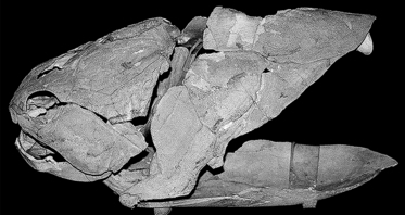
- Ahlberg PE, Trinajstic K, Long JA. The body musculature of arthrodire placoderms. Soc Vert Paleo Prog Abst. 2009;29:52A.
-
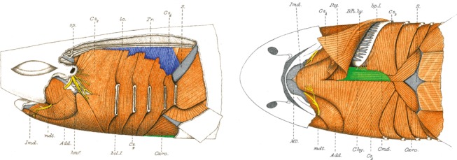
- Allis EP. The homologies of the muscles related to the visceral arches of the gnathostome fishes. Q J Microsc Sci. 1917;62:308–406.
-
- Anderson PSL. Cranial muscle homology across modern gnathostomes. Biol J Linn Soc. 2008;94:195–216.
-
- Bismuth K, Relaix F. Genetic regulation of skeletal muscle development. Exp Cell Res. 2010;316:3081–3086. - PubMed
-
- Bober E, Franz T, Arnold HH, et al. Pax-3 is required for the development of limb muscles: a possible role for the migration of dermomyotomal muscle progenitor cells. Development. 1994;120:603–612. - PubMed
Publication types
MeSH terms
LinkOut - more resources
Full Text Sources
