

Multiple precursor ion scanning of gangliosides and sulfatides with a reversed-phase microfluidic chip and quadrupole time-of-flight mass spectrometry
- PMID: 22697387
- PMCID: PMC3402638
- DOI: 10.1021/ac300254d
Multiple precursor ion scanning of gangliosides and sulfatides with a reversed-phase microfluidic chip and quadrupole time-of-flight mass spectrometry
Abstract
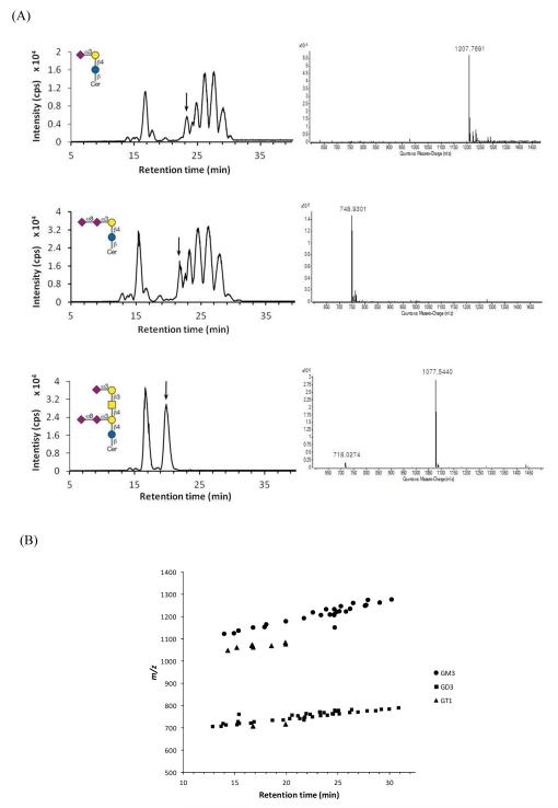
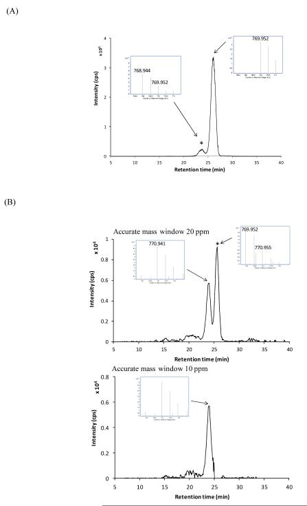
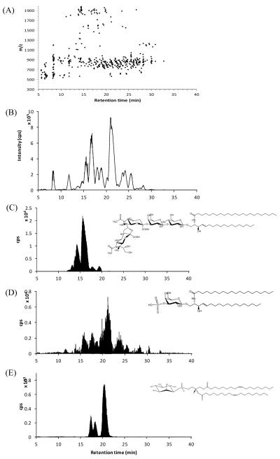
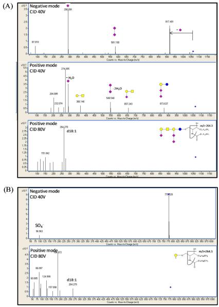
Precise profiling of polar lipids including gangliosides and sulfatides is a necessary step in understanding the diverse physiological role of these lipids. We have established an efficient method for the profiling of polar lipids using reversed-phase nano high-performance liquid chromatography microfluidic chip quadrupole time-of-flight mass spectrometry (nano-HPLC-chip Q-TOF/MS). A microfluidic chip design provides improved chromatographic performance, efficient separation, and stable nanospray while the advanced high-resolution mass spectrometer allowed for the identification of complex isobaric polar lipids such as NeuAc- and NeuGc-containing gangliosides. Lipid classes were identified based on the characteristic fragmentation product ions generated during data-dependent tandem mass spectrometry (MS/MS) experiments. Each class was monitored by a postprocessing precursor ion scan. Relatively simple quantitation and identification of intact ions was possible due to the reproducible retention times provided by the nano-HPLC chip. The method described in this paper was used to profile polar lipids from mouse brain, which was found to contain 17 gangliosides and 13 sulfatides. Types and linkages of the monosaccharides and their acetyl modifications were identified by low-energy collision-induced dissociation (CID) (40 V), and the type of sphingosine base was identified by higher energy CID (80 V). Accurate mass measurements and chromatography unveiled the degree of unsaturation and hydroxylation in the ceramide lipid tails.
Figures
References
-
- German JB, Gillies LA, Smilowitz JT, Zivkovic AM, Watkins SM. Curr. Opin. Lipidol. 2007;18:66–71. - PubMed
-
- Wenk MR. Cell. 2010;143:888–895. - PubMed
-
- Rajendran L, Simons KJ. Cell Sci. 2005;118:1099–1102. - PubMed
-
- Jacobson K, Mouritsen OG, Anderson RG. Nat. Cell. Biol. 2007;9:7–14. - PubMed
-
- Di Paolo G, De Camilli P. Nature. 2006;443:651–657. - PubMed
Publication types
MeSH terms
Substances
Grants and funding
- P42 ES02710/ES/NIEHS NIH HHS/United States
- P01 ES011269/ES/NIEHS NIH HHS/United States
- R01 ES002710/ES/NIEHS NIH HHS/United States
- R01 HD061923/HD/NICHD NIH HHS/United States
- S10RR027639/RR/NCRR NIH HHS/United States
- R01 HD059127/HD/NICHD NIH HHS/United States
- P01 ES11269/ES/NIEHS NIH HHS/United States
- 1R01HD061923/HD/NICHD NIH HHS/United States
- R01 GM049077/GM/NIGMS NIH HHS/United States
- S10 RR027639/RR/NCRR NIH HHS/United States
- R37 ES002710/ES/NIEHS NIH HHS/United States
- 5R01HD059127/HD/NICHD NIH HHS/United States
LinkOut - more resources
Full Text Sources
