. 2012 Jun 14;14(3):R94.
doi: 10.1186/bcr3210.

Dual-energy contrast-enhanced digital mammography: initial clinical results of a multireader, multicase study

Affiliations

Dual-energy contrast-enhanced digital mammography: initial clinical results of a multireader, multicase study

Clarisse Dromain et al. Breast Cancer Res. .

Abstract

Introduction: The purpose of this study was to compare the diagnostic accuracy of dual-energy contrast-enhanced digital mammography (CEDM) as an adjunct to mammography (MX) ± ultrasonography (US) with the diagnostic accuracy of MX ± US alone.

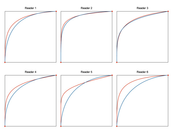
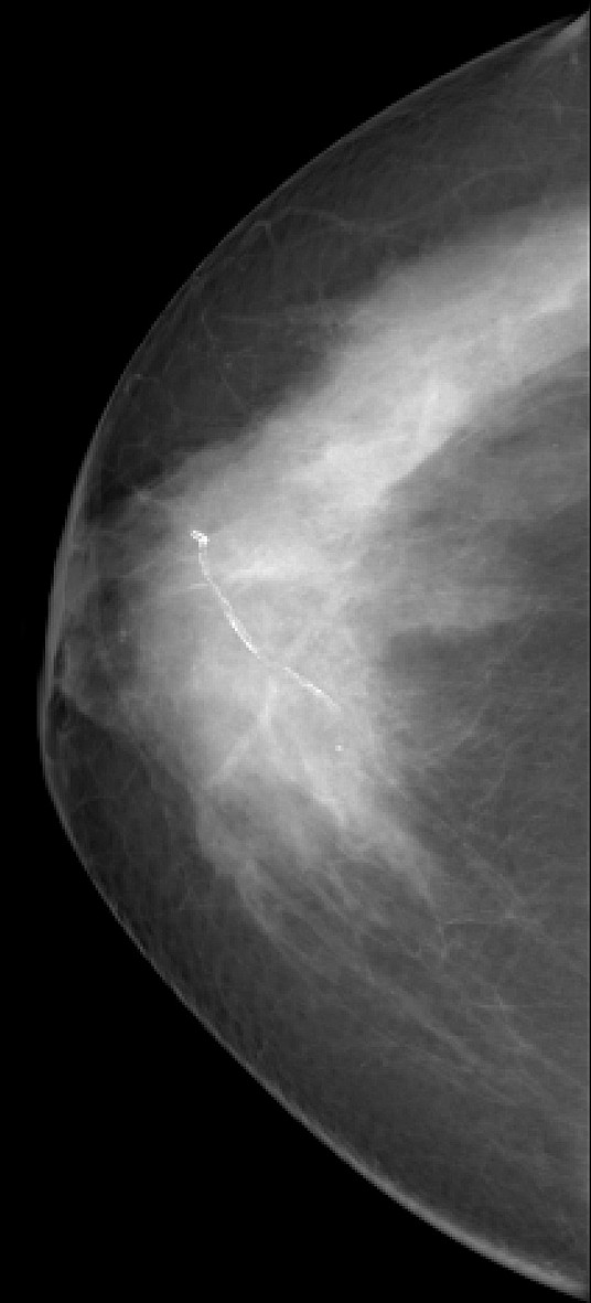
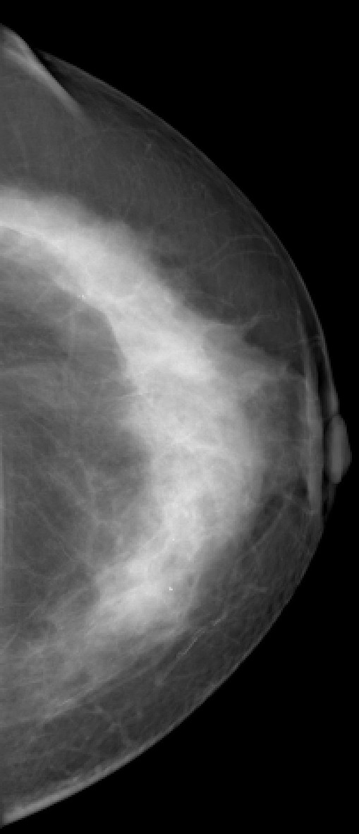
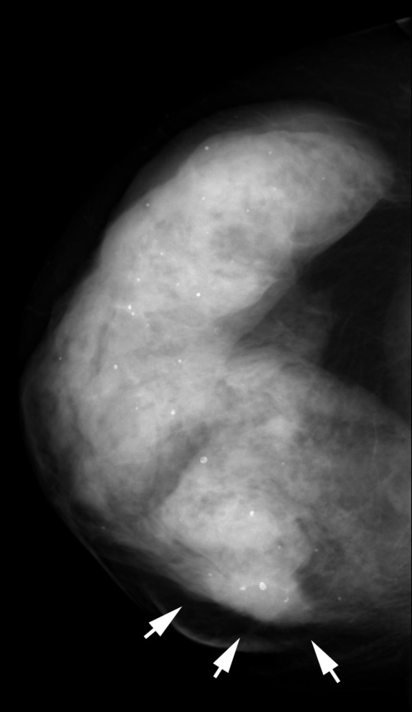
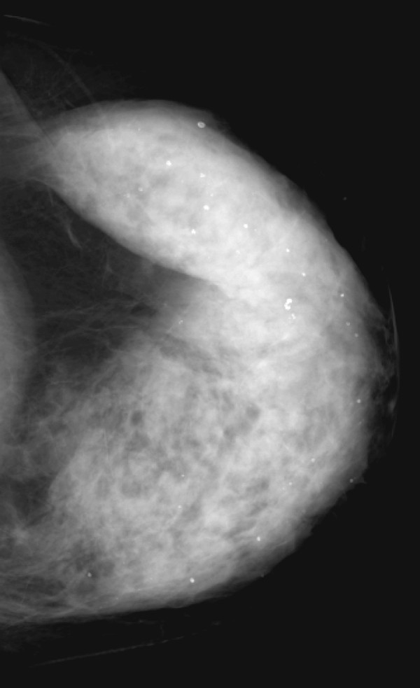
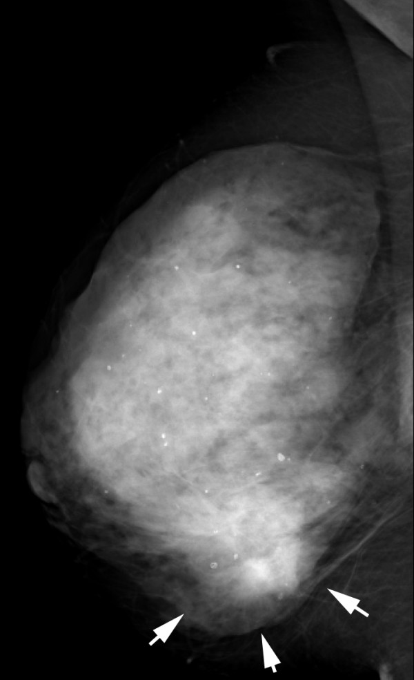
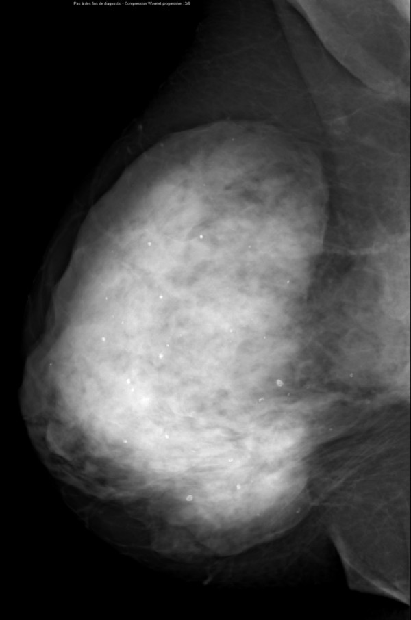
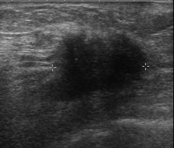
Methods: One hundred ten consenting women with 148 breast lesions (84 malignant, 64 benign) underwent two-view dual-energy CEDM in addition to MX and US using a specially modified digital mammography system (Senographe DS, GE Healthcare). Reference standard was histology for 138 lesions and follow-up for 12 lesions. Six radiologists from 4 institutions interpreted the images using high-resolution softcopy workstations. Confidence of presence (5-point scale), probability of cancer (7-point scale), and BI-RADS scores were evaluated for each finding. Sensitivity, specificity and ROC curve areas were estimated for each reader and overall. Visibility of findings on MX ± CEDM and MX ± US was evaluated with a Likert scale.

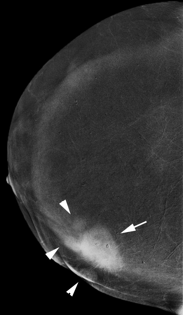
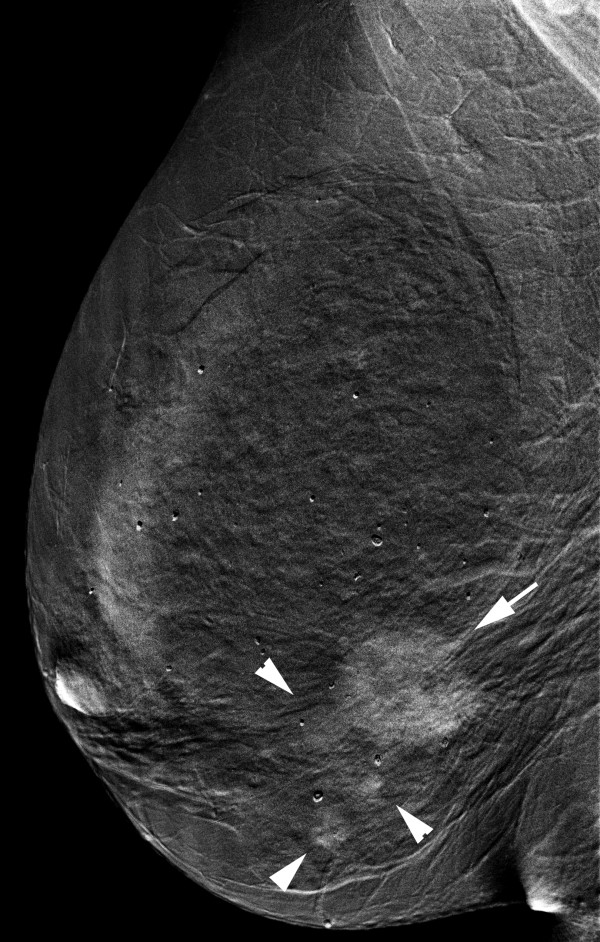
Results: The average per-lesion sensitivity across all readers was significantly higher for MX ± US ± CEDM than for MX ± US (0.78 vs. 0.71 using BIRADS, p = 0.006). All readers improved their clinical performance and the average area under the ROC curve was significantly superior for MX ± US ± CEDM than for MX ± US ((0.87 vs 0.83, p = 0.045). Finding visibility was similar or better on MX ± CEDM than MX ± US in 80% of cases.

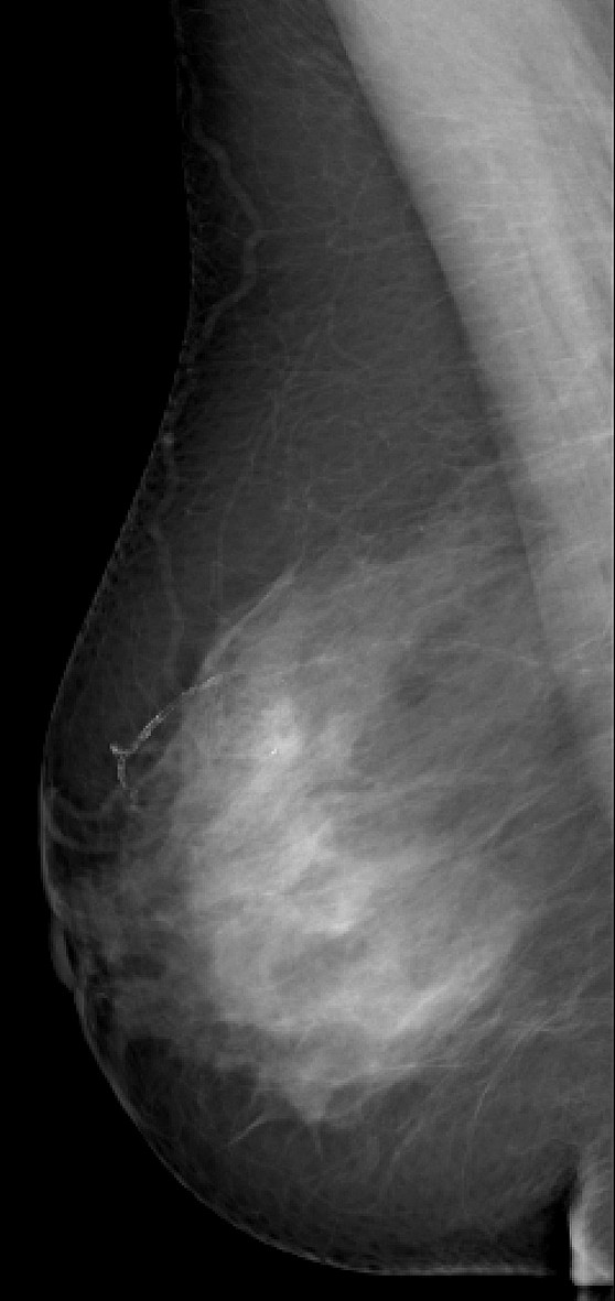
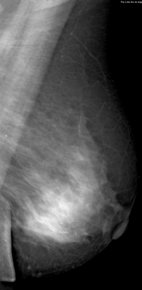
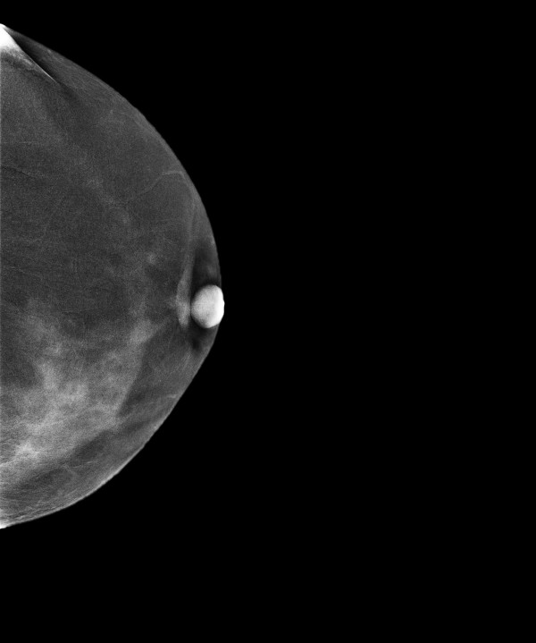
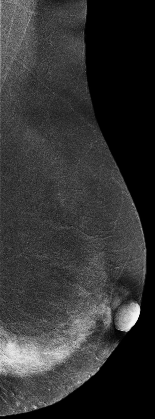
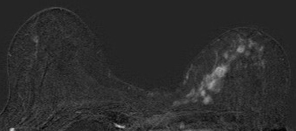
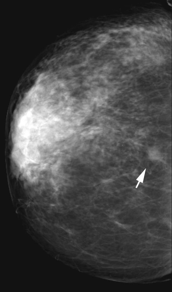
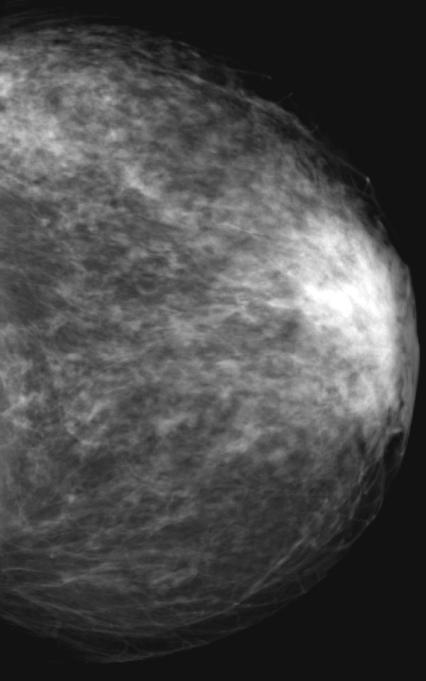
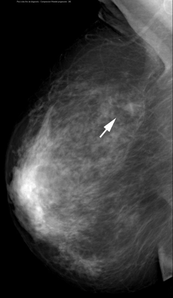
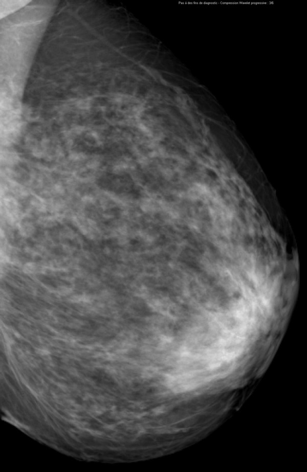
Conclusions: Dual-energy contrast-enhanced digital mammography as an adjunct to MX ± US improves diagnostic accuracy compared to MX ± US alone. Addition of iodinated contrast agent to MX facilitates the visualization of breast lesions.


Figures

Figure 1
Figure 1
Estimated average glandular dose per view (in milligrays) for contrast-enhanced digital mammography (CEDM) in comparison with mammography (MX) for 50% glandular breast. The solid line is the dose for one CEDM view, and the dotted line is for one MX view.
Figure 2
Figure 2
Estimated proper binormal receiver operating characteristic curves from BI-RADS assessments for the six readers. The red line is MX ± US ± CEDM, and the blue line is MX ± US. BI-RADS, Breast Imaging, Reporting and Data System; CEDM, contrast-enhanced digital mammography; MX, mammography; US, ultrasonography.
Figure 3
Figure 3
Invasive lobular carcinoma in a 67-year-old woman with left nipple stiffness and retraction. The right craniocaudal mammogram is normal.
Figure 4
Figure 4
Invasive lobular carcinoma in a 67-year-old woman with left nipple stiffness and retraction. The left craniocaudal mammogram shows a left cutaneous nipple and areolar thickness with no obvious lesion in the breast parenchyma.
Figure 5
Figure 5
Invasive lobular carcinoma in a 67-year-old woman with left nipple stiffness and retraction. The right mediolateral oblique mammogram is normal.
Figure 6
Figure 6
Invasive lobular carcinoma in a 67-year-old woman with left nipple stiffness and retraction. The left mediolateral oblique mammogram shows a left cutaneous nipple and areolar thickness with no obvious lesion in the breast parenchyma.
Figure 7
Figure 7
Invasive lobular carcinoma in a 67-year-old woman with left nipple stiffness and retraction. The iodine-enhanced, contrast-enhanced digital mammography, craniocaudal image clearly depicts non-mass regional enhancement in the inner quadrant.
Figure 8
Figure 8
Invasive lobular carcinoma in a 67-year-old woman with left nipple stiffness and retraction. The iodine-enhanced, contrast-enhanced digital mammography, mediolateral oblique images clearly depicts non-mass regional enhancement in the inferior quadrant.
Figure 9
Figure 9
Invasive lobular carcinoma in a 67-year-old woman with left nipple stiffness and retraction. The contrast-enhanced transaxial breast magnetic resonance image shows the same non-mass regional enhancement than CEDM images.
Figure 10
Figure 10
Multifocal invasive ductal carcinoma in a 53-year-old woman with dense breasts. The right craniocaudal view mammogram shows a very dense breast with an uncertain opacity in the inner quadrant (arrows).
Figure 11
Figure 11
Multifocal invasive ductal carcinoma in a 53-year-old woman with dense breasts. The left craniocaudal view mammogram shows a very dense breast with no obvious lesion.
Figure 12
Figure 12
Multifocal invasive ductal carcinoma in a 53-year-old woman with dense breasts. The right mediolateral oblique view mammogram shows a very dense breast with an uncertain opacity in the inferior quadrant (arrows).
Figure 13
Figure 13
Multifocal invasive ductal carcinoma in a 53-year-old woman with dense breasts. The left mediolateral oblique view mammogram shows a very dense breast with no obvious lesion.
Figure 14
Figure 14
Multifocal invasive ductal carcinoma in a 53-year-old woman with dense breasts. The ultrasound image clearly demonstrates a 26-mm hypoechoic mass with irregular margins highly suggestive of malignancy.
Figure 15
Figure 15
Multifocal invasive ductal carcinoma in a 53-year-old woman with dense breasts. The iodine-enhanced, contrast-enhanced digital mammography, right craniocaudal image readily depicts the main mass (arrow) and three additional adjacent nodules (arrowheads).
Figure 16
Figure 16
Multifocal invasive ductal carcinoma in a 53-year-old woman with dense breasts. The iodine-enhanced, contrast-enhanced digital mammography, right mediolateral oblique image readily depict the main mass (arrow) and three additional adjacent nodules (arrowheads).
Figure 17
Figure 17
Histologically proven normal breast parenchyma in a 69-year-old woman. The right craniocaudal view mammogram shows an opacity with irregular margins (arrow).
Figure 18
Figure 18
Histologically proven normal breast parenchyma in a 69-year-old woman. The left craniocaudal view mammogram is normal.
Figure 19
Figure 19
Histologically proven normal breast parenchyma in a 69-year-old woman. The right mediolateral oblique view mammogram shows an opacity with irregular margins (arrow). This lesion is classified as a BI-RADS (Breast Imaging, Reporting and Data System) score of 4.
Figure 20
Figure 20
Histologically proven normal breast parenchyma in a 69-year-old woman. The left mediolateral oblique view mammogram is normal.
Figure 21
Figure 21
Histologically proven normal breast parenchyma in a 69-year-old woman. The iodine-enhanced, contrast-enhanced digital mammography, right craniocaudal image clearly demonstrates no obvious contrast uptake in the area of the nodule depicted on mammography.
Figure 22
Figure 22
Histologically proven normal breast parenchyma in a 69-year-old woman. The iodine-enhanced, contrast-enhanced digital mammography, mediolateral oblique right image clearly demonstrates no obvious contrast uptake in the area of the nodule depicted on mammography. A core-needle stereotactic biopsy confirmed that the opacity was normal glandular tissue.

References

    1. Humphrey LL, Helfand M, Chan BK, Woolf SH. Breast cancer screening: a summary of the evidence for the US Preventive Services Task Force. Ann Intern Med. 2002;137:347–360. - PubMed
    1. Rosenberg RD, Hunt WC, Williamson MR, Gilliland FD, Wiest PW, Kelsey CA, Key CR, Linver MN. Effects of age, breast density, ethnicity, and estrogen replacement therapy on screening mammographic sensitivity and cancer stage at diagnosis: review of 183,134 screening mammograms in Albuquerque, New Mexico. Radiology. 1998;209:511–518. - PubMed
    1. Kolb T, Lichy J, Newhouse J. Comparison of the performance of screening mammography, physical examination, and breast US and evaluation of factors that influence them: an analysis of 27,825 patient evaluations. Radiology. 2002;225:165–175. doi: 10.1148/radiol.2251011667. - DOI - PubMed
    1. Bird RE, Wallace TW, Yankaskas BC. Analysis of cancer missed at screening mammography. Radiology. 1992;184:613–617. - PubMed
    1. Pisano ED, Gatsonis C, Hendrick E, Yaffe M, Baum JK, Acharyya S, Conant EF, Fajardo LL, Bassett L, D'Orsi C, Jong R, Rebner M. Digital Mammographic Imaging Screening Trial (DMIST) Investigators Group. Digital Mammographic Imaging Screening Trial (DMIST) Investigators Group. Diagnostic performance of digital mammography versus film mammography for breast-cancer screening. N Engl J Med. 2005;353:1773–1783. doi: 10.1056/NEJMoa052911. - DOI - PubMed

Publication types